Immune signals’ links to breast aging shift across menopause
In 81 women with biopsies 2–10 years apart, the same immune-related gene patterns were linked to slower breast tissue shrinkage across menopause but faster shrinkage afterward.
Our read
Mechanisms- Studied:
- Cohort study of 81 women with paired benign breast biopsies, 2-10 years
- What changed:
- Higher immune signals were linked to slower breast lobule shrinkage during the menopausal transition, but faster shrinkage after menopause
- The catch:
- Registry-reported menopause age can misclassify women near the menopausal boundary
- Stake:
- An author holds patents licensed to Unity Biotechnology.
- Why it’s aging research:
- It studies something that builds up in the body with age.
Editor of record: Boris Djordjevic · AgingResearch.News
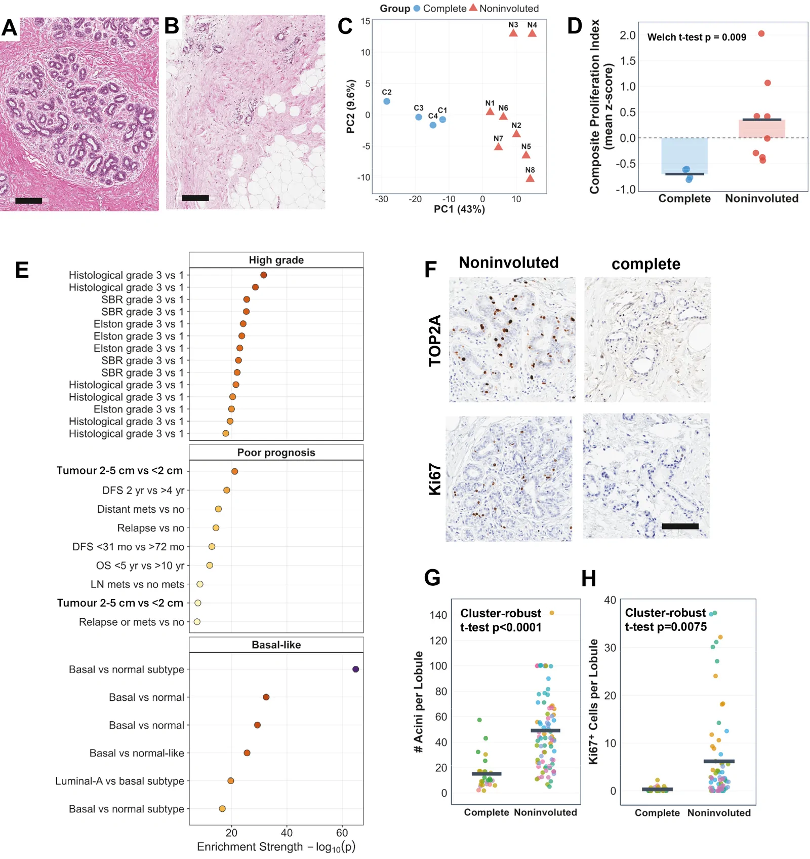
An observational study followed 81 women aged 45–55 at their first noncancerous breast biopsy, with a second biopsy 2–10 years later. Researchers measured gene activity and shrinkage of lobules, clusters of milk-producing glands.
Gene activity related to inflammatory signals from senescent cells—cells that stop dividing—and the body's built-in immune defenses showed opposite associations with shrinkage. These patterns were linked to slower shrinkage across the menopausal transition but faster shrinkage after menopause. Imaging in small subsets linked stalled shrinkage around menopause to immune cells positioned apart from cells carrying a protein associated with senescence. After menopause, a pattern associated with cell removal featured immune cells contacting those marked cells. The authors concluded that interpreting these biological signals should account for menopausal timing.
Why it matters
Incomplete shrinkage of breast lobules is associated with higher breast cancer risk. Understanding its relationship with immune activity may help researchers interpret tissue signals in the context of menopause.
Caveats
The observational design could not show that immune-cell contact caused cell removal or faster shrinkage. Imaging involved 14–16 women at baseline and 14 at follow-up; the findings concern noncancerous breast tissue.
From the paper
- Participants
We analysed 81 women with paired benign biopsies (baseline age 45–55; follow-up 2–10 years) who had noninvoluted lobules at baseline.
Quoted word for word from the full text on PubMed Central.
- Cellular senescence
- Immune response
- Senescence-associated secretory phenotype (SASP)
- Breast cancer
- CD16+ innate-effector engagement
- Humans
The paper
Mayo Clinic in Florida
EBioMedicine, Aug 31, 2026, Open access

Comments
Be the first to comment.