Age-related decline of platelet-derived growth factor receptor alpha in mesenchyme surrounding meibomian gland leads to meibomian gland dysfunction
Abstract
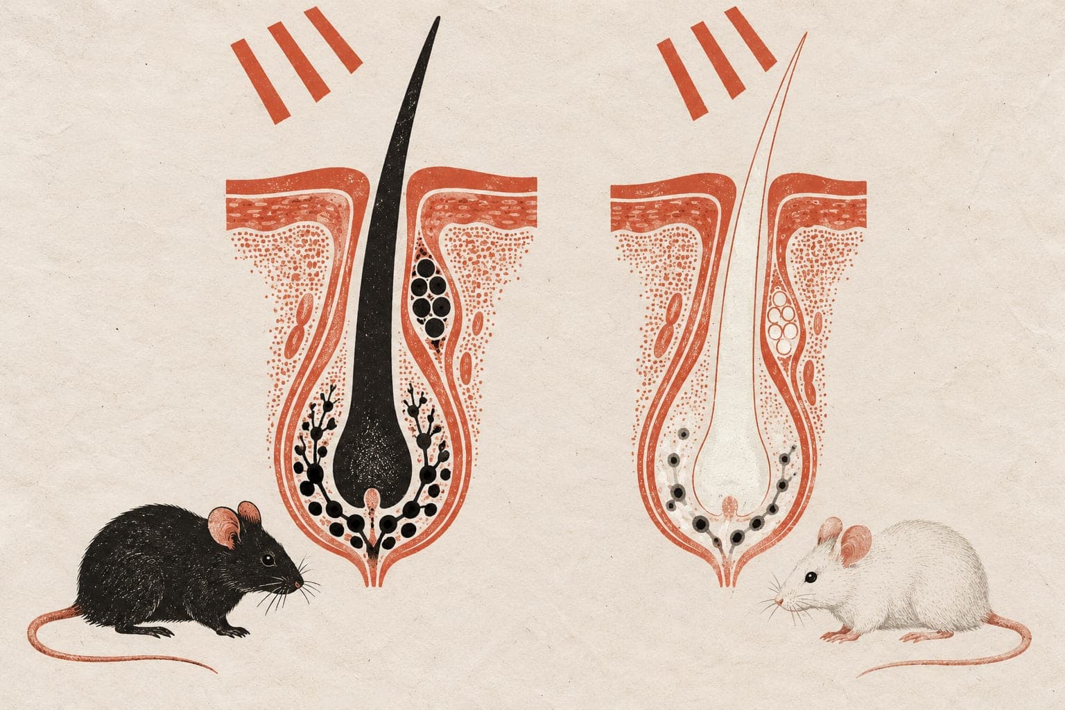
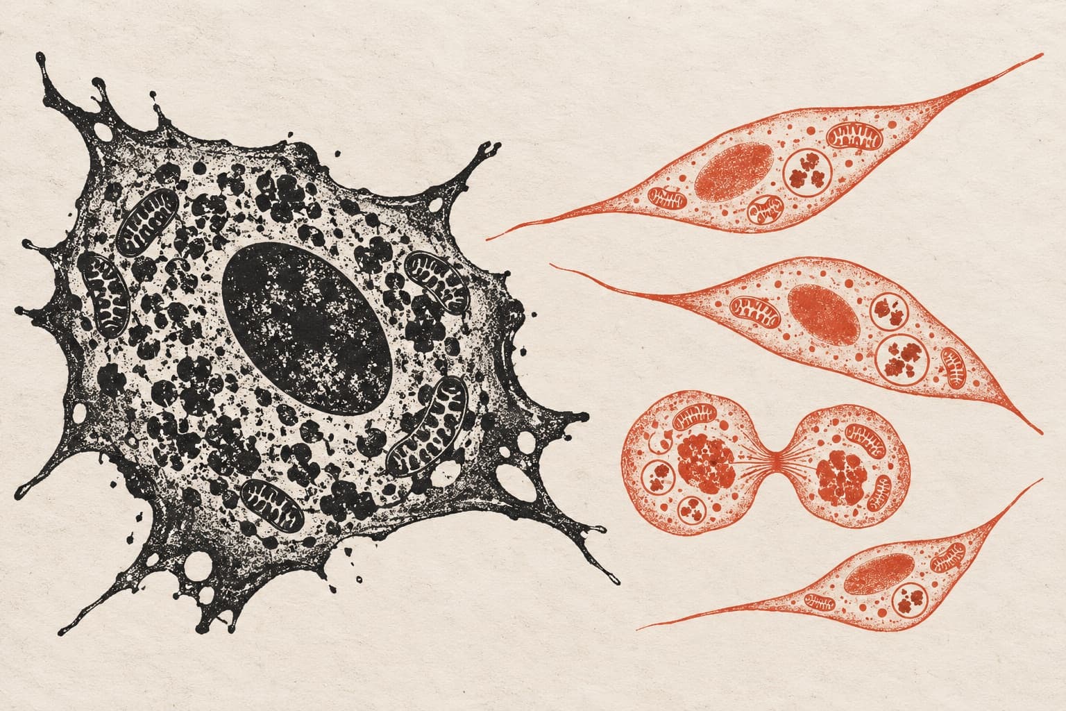
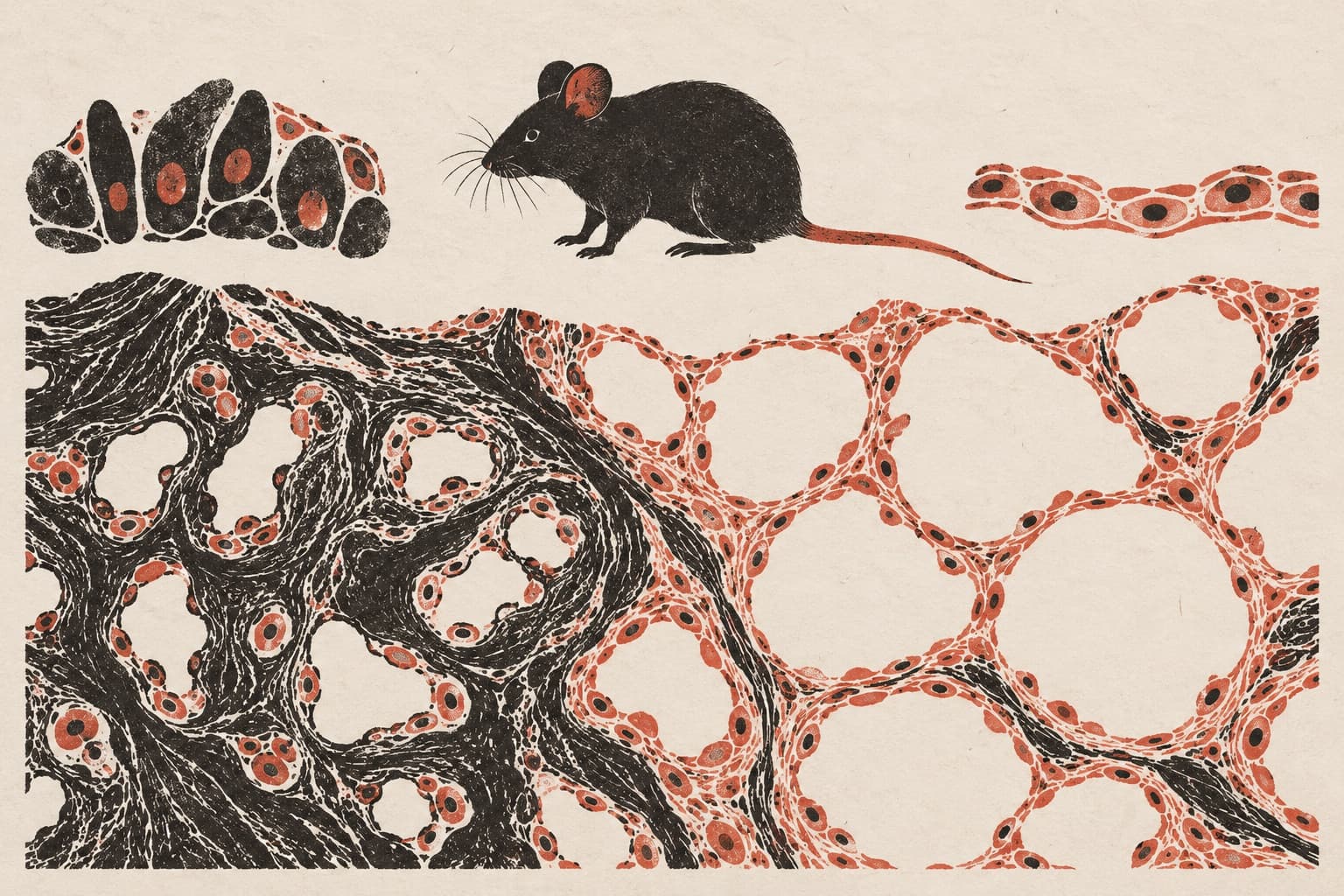
The meibomian glands (MGs) in the eyelids produce oil to prevent tear evaporation, a function that declines with age. The mechanisms underlying MGs homeostasis and the pathogenesis leading to age-related meibomian gland dysfunction (ARMGD) remain largely unexplored. MGs contain secretory acini enveloped by a substantial mesenchyme. Previous studies have identified that ARMGD resulted from the failure to replenish acinar cells due to their senescence. Few studies, however, explored the role of age-related changes in mesenchyme contributing to ARMGD. Here, we demonstrated that an age-dependent decline in platelet-derived growth factor receptor alpha (PDGFR-α) within the peri-glandular mesenchyme was associated with ARMGD progression in both mice and humans. Furthermore, we discovered PDGFR-α⁺ cells co-expressing hybrid mesenchymal-epithelial markers in mesenchyme adjacent to MGs. This led us to hypothesize the existence of a plastic cell population whose function was impaired by aging. In MGs of young mice, inhibiting PDGFR-α expression upregulated cellular senescence genes (p16INK4a and p21Cip1/Waf1) and downregulated epithelial marker gene (krt14) within the mesenchyme, indicating that PDGFR-α downregulation induced mesenchymal senescence and plasticity losing. Additionally, PDGFR-α inhibition in young mouse MGs reduced acinar cell count and proliferation (ki67 expression), culminating in MGs atrophy and evaporative dry eye. Our findings highlighted the significance of mesenchymal-epithelial interaction in maintaining MGs homeostasis and proposed PDGFR-α-signaling within mesenchyme as a potential therapeutic target for ARMGD.
- PDGFRA
- Cell cycle regulation
- Cellular homeostasis
- Meibomian gland dysfunction
- Age-related meibomian gland dysfunction
- Humans
The paper
Nanfang Hospital
Experimental Eye Research, 25 Mar 2026