Largely Distinct Post-Translational Modifications Differentiate Skeletal Muscle Wasting Caused by Cancer, Dexamethasone and Aging
Abstract
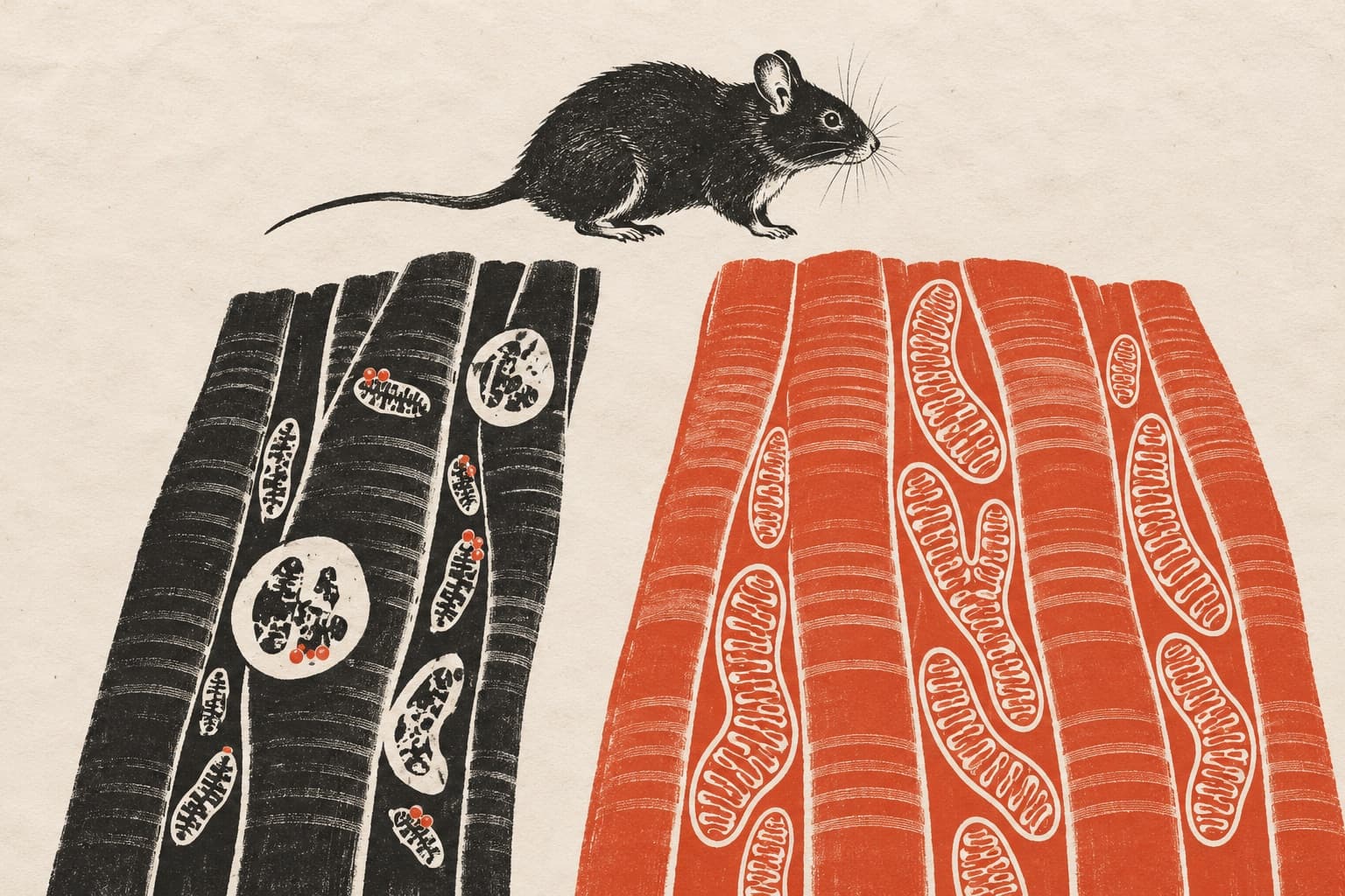
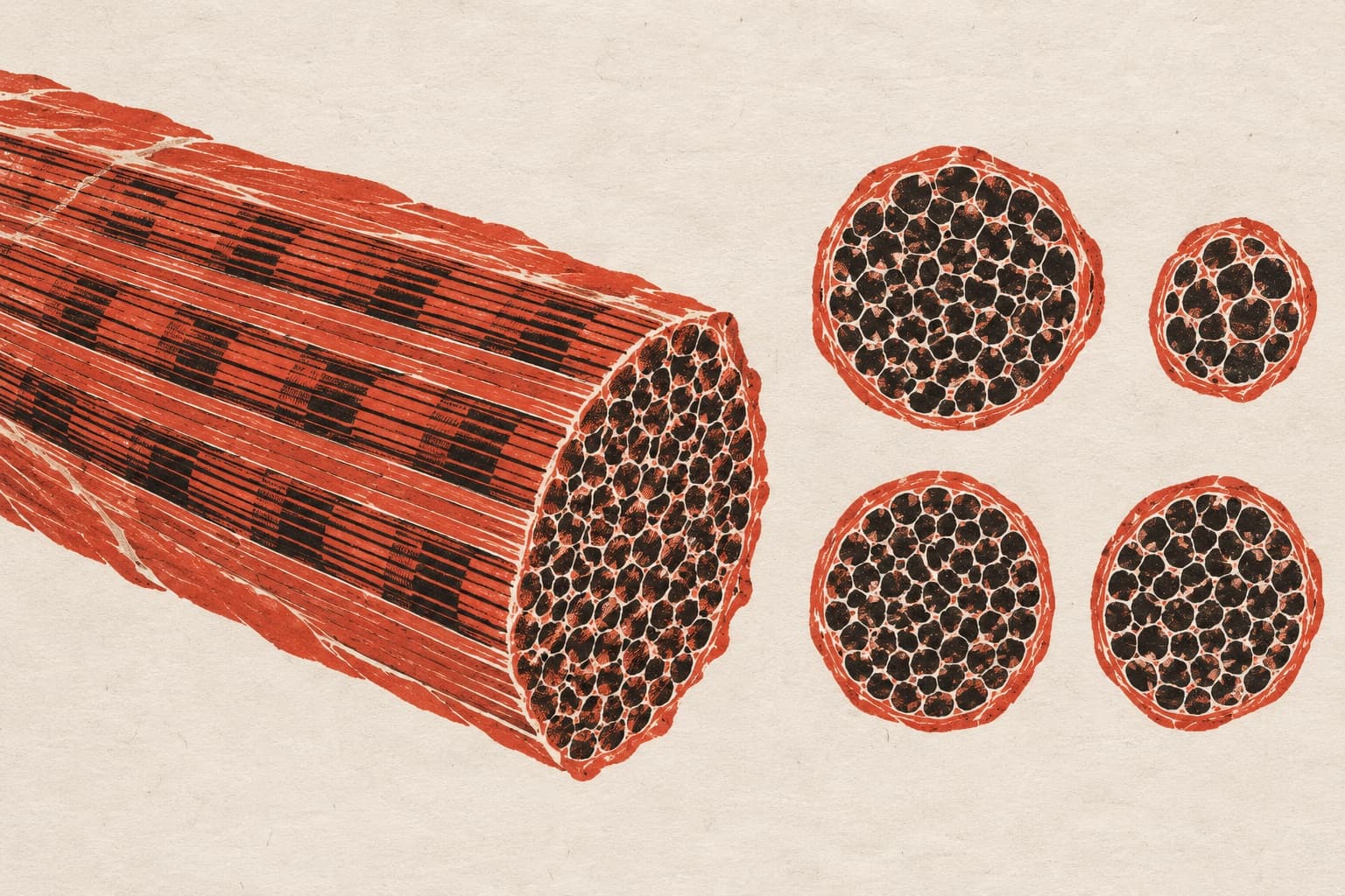
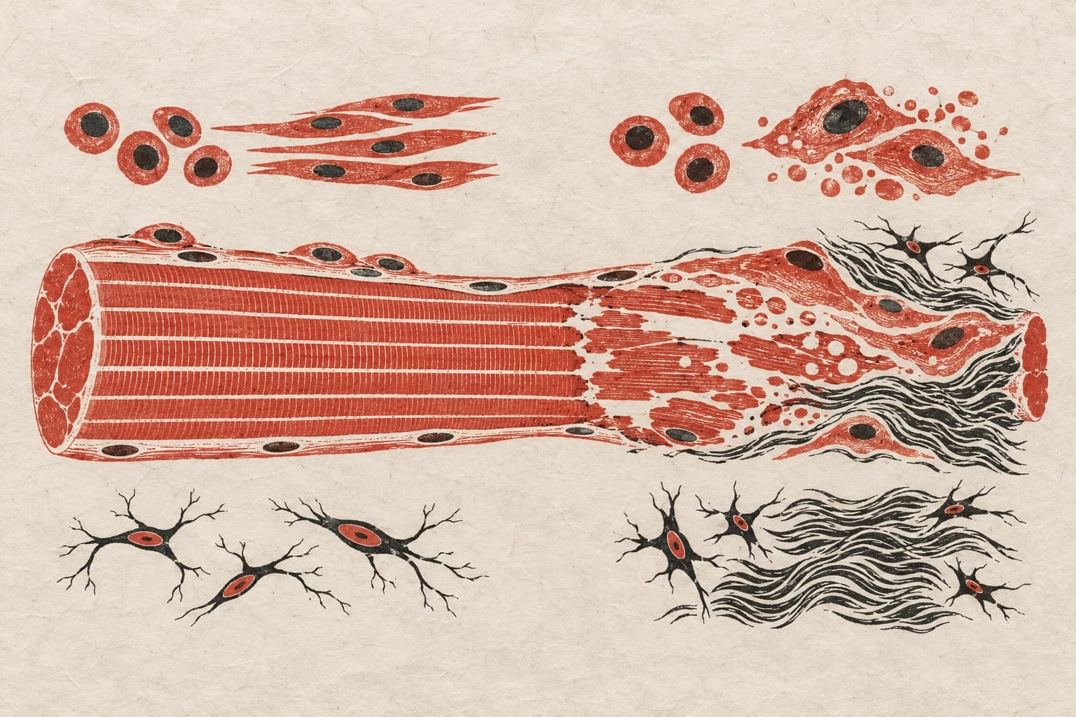
BACKGROUND: Skeletal muscle wasting and weakness are prominent disease features. Originally considered to arise from common transcriptional changes, recent analyses demonstrated that different stimuli induce muscle wasting via largely distinct mRNA and protein changes. METHODS: Here, we examined the post-translational modifications (PTMs) associated with muscle wasting induced by cancer (n = 15 078), dexamethasone (n = 15 078) and aging (n = 8777) in mice by utilising the JUMPptm pipeline to recover modified peptides from TMT (tandem mass tag) mass spectrometry analyses. RESULTS: We find that most PTMs that are significantly regulated are stimulus-specific and that only a few are cross-shared (n = 10; p < 0.05). These include P27 dihydroxylation of Lrpprc (leucine-rich pentatricopeptide repeat containing), an RNA binding protein and transcriptional co-activator mutated in Leigh syndrome, a mitochondrial disease. Contrary to the stimulus-specificity of other atrophy-associated PTMs, P27 dihydroxylation of Lrpprc declines (~20%; p < 0.05) with muscle wasting irrespective of the atrophic trigger. Electroporation of dihydroxylation-resistant LrpprcP27A (which mimics the reduction in Lrpprc dihydroxylation that occurs with atrophy) reduces muscle force in young (~23%-39%; p < 0.01) and old (~26%-36%; p < 0.01) male mice compared to the contralateral electroporation of LrpprcWT, indicating that a decline in Lrpprc P27 dihydroxylation contributes to muscle weakness in response to diverse catabolic stimuli. Comparison of LrpprcWT versus GFP electroporation indicates that there are mostly non-significant effects (p > 0.05) on muscle force in young and old mice. Mechanistically, LrpprcP27A does not affect proteostasis and mitochondrial function compared to control LrpprcWT but impairs (> 60% decline; p < 0.05) the expression of genes necessary for muscle strength, including the apelin receptor Aplnr and Col6a2/6 collagens. Moreover, LrpprcP27A reduces type 2b myofibre size (13% decline; p < 0.01) in old but not in young age. CONCLUSIONS: These analyses identify atrophy-associated PTMs that provide refined biomarkers for fingerprinting the atrophic stimulus. Although most PTMs are stimulus-specific, P27 dihydroxylation of Lrpprc declines during muscle wasting induced by cancer, dexamethasone and aging, suggesting that this is a general atrophy marker. Experimental up-regulation of the atrophy-mimicking variant LrpprcP27A reduces muscle force compared to wild-type Lrpprc in young and old mice, suggesting that atrophy-associated P27 dihydroxylation contributes to disease-associated muscle weakness.
The paper
St. Jude Children's Research Hospital
Journal of Cachexia, Sarcopenia and Muscle, 1 Feb 2026