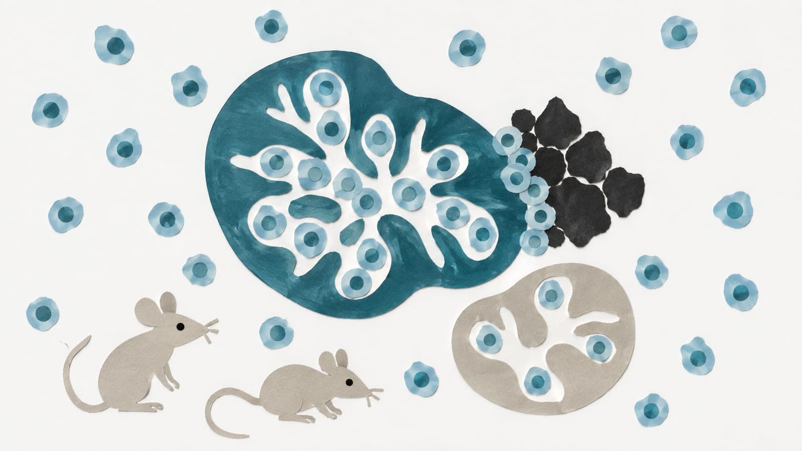
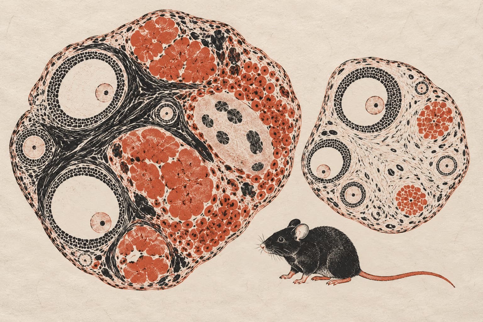
Mechanical Insights into the Distinct Effects of Ovariectomy Versus Adrenalectomy on Age-Related Thymic Atrophy in Female Mice
Abstract
Age-related thymic atrophy, a hallmark of immunosenescence linked to age-related diseases, involves gonadal and adrenal steroid hormones, but their distinct roles and mechanisms in this process remain unclear. Through biochemical, histological, and RNA-seq analyses, we comprehensively explored the mechanisms underpinning age-related thymic atrophy in response to ovariectomy (OVX) versus adrenalectomy (ADX) in female mice. Compared to the sham controls, OVX overtly ameliorated age-related thymic atrophy, as evidenced by increased thymus mass, a larger gross thymus area, and denser cortex cellularity. In contrast, ADX evidently accelerated age-related thymic atrophy, characterized by increased adipose infiltration and decreased cortex/medulla ratio, obscure cortico-medullary junctions, and sparser thymic cortical cells. Unexpectedly, combined OVX and ADX displayed a more pronounced effect than OVX alone in ameliorating age-related thymic atrophy. Mechanistically, OVX decreased while ADX increased the circulating 17β-estradiol levels in female mice, which drove these opposing outcomes potentially by promoting Pparg-mediated thymic fat deposition and blocking Cdk1-dependent thymocyte cell cycle progression. Although OVX eliminated gonadal 17β-estradiol production, it appeared to trigger a compensatory adrenal-dependent estrogen biosynthesis, whereas combined OVX and ADX nearly eliminated all estrogen sources, thus leading to a more pronounced effect than OVX alone in ameliorating age-related thymic atrophy in female mice. Notably, OVX increased while ADX decreased serum corticosterone levels, but these alterations exerted minimal impacts on age-related thymic atrophy, highlighting a pivotal role of estrogens over glucocorticoids in accelerating age-related thymic atrophy in females. Undesirably, although OVX ameliorated age-related thymic atrophy, it appeared to simultaneously increase autoimmune susceptibility by downregulating thymic Cd74 expression. Taken together, our results indicate that OVX ameliorates while ADX accelerates age-related thymic atrophy in females. Estrogens rather than glucocorticoids act as the predominant regulator of this process, potentially via promoting Pparg-dependent fat deposition and blocking Cdk1-dependent thymocyte cycle progression. However, OVX-induced estrogen depletion also elevated autoimmune risk, emphasizing the need to balance benefits and risks in regulating thymic aging.
The paper
Sichuan Agricultural University
International Journal of Molecular Sciences, 20 Jan 2026