Role of capillary rarefaction in age-related changes of external urethral sphincter muscle of female mice
Abstract
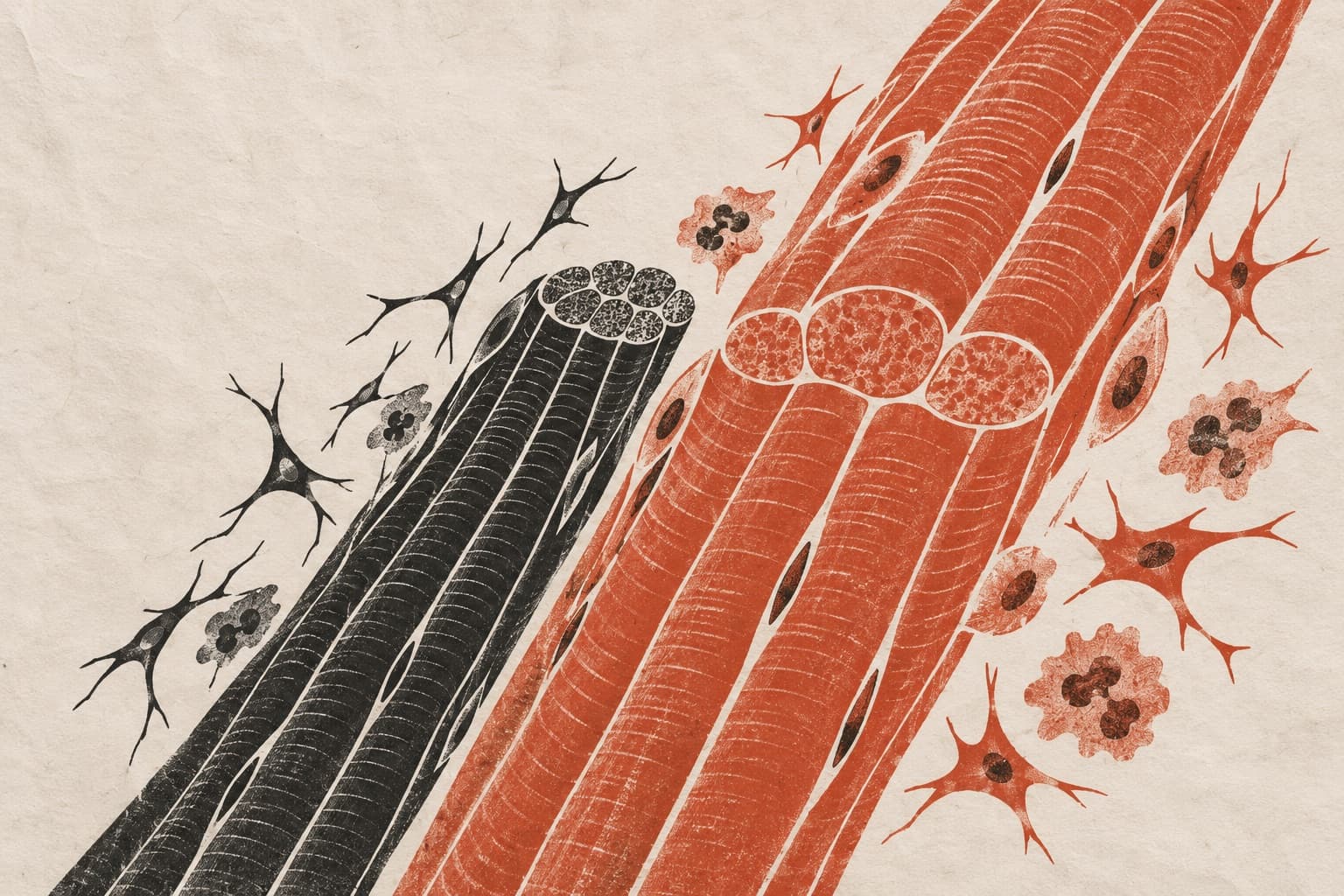
Age-related loss of volume and strength of the external urethral sphincter (EUS) is involved in the pathogenesis of stress urinary incontinence. Here, we explored if aged EUS undergoes capillary rarefaction, a characteristic of sarcopenia. Ca²⁺ dynamics in pericytes of EUS capillaries were also investigated to understand their roles in regulating EUS blood supply. Florescence immunohistochemistry was employed to compare the density of striated muscle fibres, fibroblasts, capillaries and pericytes between young and aged female mouse EUS. Electrical filed stimulation (EFS)-induced Ca²⁺ dynamics in EUS capillary pericytes were investigated using young NG2-GCaMP6 mice. In comparison to young EUS, aged EUS displayed a reduced density of striated muscle fibres, an increased density of PDGFRα⁺ fibroblasts, as well as a reduced density of capillaries and pericytes. EFS evoked prolonged Ca²⁺ transients and transient Ca²⁺ reductions in α-smooth muscle actin negative pericytes. The EFS-induced Ca²⁺ transients were prevented by either guanethidine (10 µM) or phentolamine (1 µM), despite the lack of pericapillary tyrosine hydroxylase-positive sympathetic fibres. The EFS-induced Ca²⁺ reductions were prevented by L-nitroarginine (10 µM). Pericytes were not innervated by neuronal NOS (nNOS)-positive nerve fibres, while EUS muscle fibres express sarcolemmal nNOS immunoreactivity. Thus, capillary rarefaction may contribute to the age-related EUS loss associated fibrosis. The Ca²⁺ reductions in pericytes appear to be mediated by NO released from EUS muscle fibres. Since pericytes express ANO1 Ca²⁺-activated chloride channels, the Ca²⁺ reductions would cause hyperpolarisation that spreads to the upstream resulting in arteriolar dilatation to increase blood supply to contracting EUS fibres.
The paper
Nagoya City University
Cell and Tissue Research, 28 Nov 2025