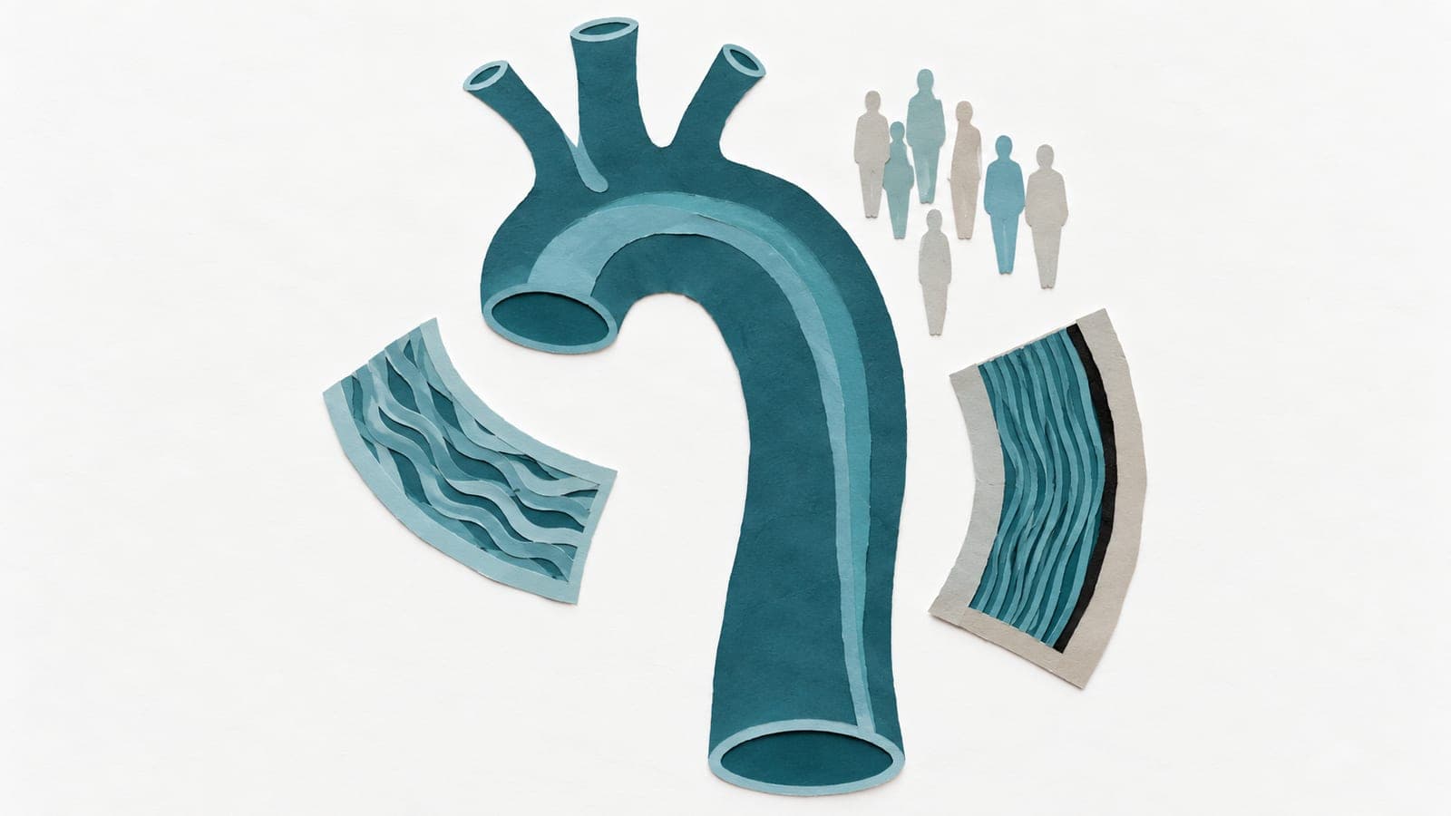
Aortic flow is associated with aging and exercise capacity
Abstract
AIMS: Increased blood flow eccentricity in the aorta has been associated with aortic (AO) pathology, however, its association with exercise capacity has not been investigated. This study aimed to assess the relationships between flow eccentricity parameters derived from 2-dimensional (2D) phase-contrast (PC) cardiovascular magnetic resonance (CMR) imaging and aging and cardiopulmonary exercise test (CPET) in a cohort of healthy subjects. METHODS AND RESULTS: One hundred and sixty-nine healthy subjects (age 44 ± 13 years, M/F: 96/73) free of cardiovascular disease were recruited in a prospective study (NCT03217240) and underwent CMR, including 2D PC at an orthogonal plane just above the sinotubular junction, and CPET (cycle ergometer) within one week. The following AO flow parameters were derived: AO forward and backward flow indexed to body surface area (FFi, BFi), average flow displacement during systole (FDsavg), late systole (FDlsavg), diastole (FDdavg), systolic retrograde flow (SRF), systolic flow reversal ratio (sFRR), and pulse wave velocity (PWV). Exercise capacity was assessed by peak oxygen uptake (PVO₂) from CPET. The mean values of FDsavg, FDlsavg, FDdavg, SRF, sFRR, and PWV were 17 ± 6%, 19 ± 8%, 29 ± 7%, 4.4 ± 4.2 mL, 5.9 ± 5.1%, and 4.3 ± 1.6 m/s, respectively. They all increased with age (r = 0.623, 0.628, 0.353, 0.590, 0.649, 0.598, all P < 0.0001), and decreased with PVO₂ (r = -0.302, -0.270, -0.253, -0.149, -0.219, -0.161, all P < 0.05). A stepwise multivariable linear regression analysis using left ventricular ejection fraction (LVEF), FFi, and FDsavg showed an area under the curve of 0.769 in differentiating healthy subjects with high-risk exercise capacity (PVO₂ ≤ 14 mL/kg/min). CONCLUSION: AO flow haemodynamics change with aging and predict exercise capacity. REGISTRATION: NCT03217240.
The paper
National Heart Centre Singapore; National University of Singapore
European Heart Journal Open, 26 Aug 2023, Open access