Protein loss altered kidney filter structure in mice
Mice engineered to lack a protein developed abnormal kidney-filter deposits, but the loss did not severely impair kidney function.
Limit: Direct and indirect causes of mesangial expansion were not distinguished
Edited by Boris Djordjevic · AgingResearch.News
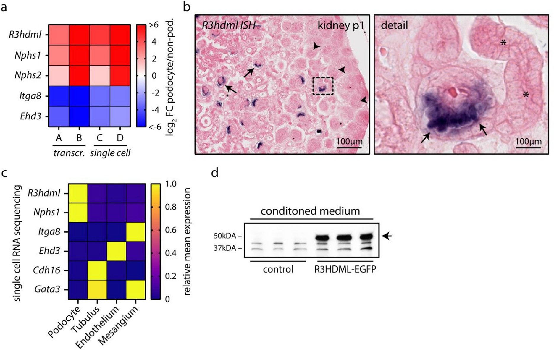
In experiments in mice, researchers deleted the gene for R3HDML, a protein made and released by podocytes, cells that form part of the kidney’s filters. The mice developed hump-like deposits of collagen IV, a structural protein, in the glomerular basement membrane, a supporting layer of those filters. They also developed expansion of the tissue between the filters’ small blood vessels.
The deposits caused the podocytes’ extensions to retract and their internal structural fibers to form clusters. Despite these changes, loss of the protein did not severely impair kidney function during aging, diabetes-related kidney disease, or injury caused by interrupting and restoring blood flow. The findings identified a role for R3HDML in maintaining kidney-filter structure.
Why it matters
The work addresses how kidney-filtering cells preserve their supporting layer during aging and diabetes-related kidney disease.
Caveats
These gene-deletion experiments were in mice and do not establish whether the protein has the same role in people.