A gene primes mouse retinal cells to divide and make neurons
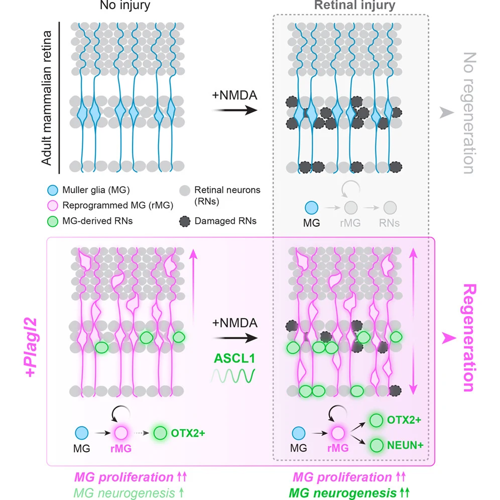
In adult mice, Plagl2 reprogrammed retinal support cells to divide and gain neuron-making potential, which retinal injury further enhanced.
Our read
Interventions- Studied:
- Experiments in adult mouse retinal Müller glia
- What changed:
- Plagl2 made adult mouse retinal support cells divide and gain neuron-making potential
- The catch:
- Neuron-making potential, rather than lost-neuron replacement or improved vision, was reported
- In the record:
- One of 6 papers on N-methyl-D-aspartate on record: none yet with results in people, 3 in animals.
- Why it’s aging research:
- Repairing, replacing or regenerating the aging body.
Editor of record: Boris Djordjevic · AgingResearch.News
Adult mouse Müller glia—support cells in the eye’s light-sensing retina—were reprogrammed in animal experiments using Plagl2, a gene that controls other genes’ activity. The cells entered a precursor-like state, combining the ability to multiply with the potential to become neurons.
The researchers examined tissue, recorded time-lapse images and measured gene activity in individual cells. Plagl2 prompted regulated rounds of re-entry into the cell division cycle. Retinal injury caused by a chemical called N-methyl-D-aspartate further increased the cells’ potential to become neurons. That potential favored neuron types found in a retinal layer called the inner nuclear layer.
Why it matters
Lost retinal neurons are a target for regeneration using cells already present in adult tissue. The authors suggest that adapting reprogramming strategies across cell types could support broader approaches to tissue regeneration.
Caveats
The abstract reports neuron-making potential, not whether lost neurons were replaced or vision improved. The experiments were in adult mice, not people.
Comments
Be the first to comment.