Measures of aging in cell structures linked to disease risk
Among 61,931 people, four or more prematurely aged compartments were linked to up to 22-fold higher kidney disease risk and up to 8-fold higher mortality risk.
Cohort study
The catch: Fluid proteins served as proxies for organelle aging rather than direct measurements inside cells
Edited by Boris Djordjevic · AgingResearch.News
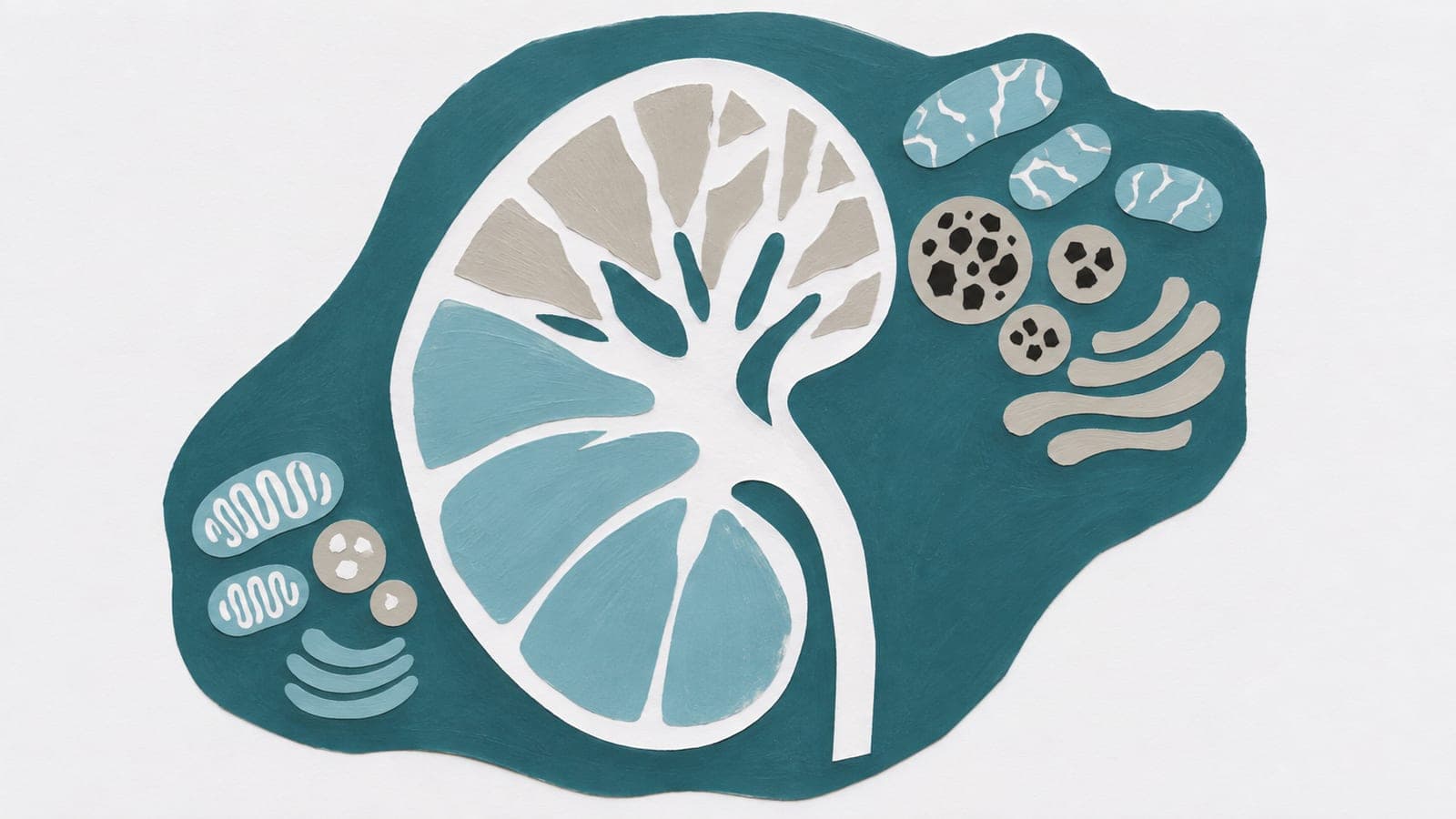
Researchers analyzed protein measurements from 61,931 people in an observational study. The proteins came from blood plasma and cerebrospinal fluid, the fluid around the brain and spinal cord. They used proteins enriched in particular organelles—specialized structures within cells—to model the aging of 10 organelles across the human lifespan.
The estimated aging patterns differed among organelles. Estimates for lysosomes, which break down cellular waste, cells’ outer membranes and the Golgi apparatus, which processes and packages proteins, accelerated rapidly after age 60. More compartments with accelerated estimated aging were linked to higher disease risk. Different major diseases also had distinct patterns of estimated organelle aging.
Why it matters
Aging does not affect every cell structure alike. Measuring these differences may help researchers investigate why people vary in their susceptibility to disease.
Caveats
The observational analysis used protein-based estimates of organelle aging, so it cannot establish cause and effect. The preprint has not been peer-reviewed; its abstract does not specify the risk comparison groups or follow-up.
The paper
Organelle Aging Predicts Human Disease
Stanford University
bioRxiv, Nov 3, 2026, Open access, CC BY-NC-ND, Preprint, not yet peer-reviewed


Comments
Be the first to comment.