Imaging method measures individual telomeres in DNA samples
The assay measured telomeres spanning about 50 base pairs to more than 50 kilobases within the same DNA sample.
Limit: The abstract omits validation sample sizes
Edited by Boris Djordjevic · AgingResearch.News
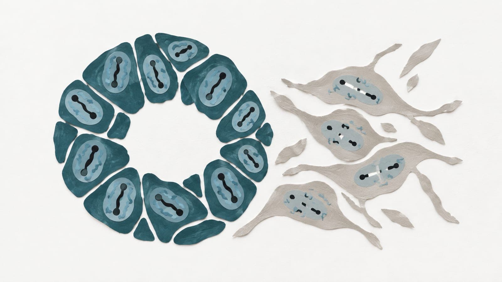
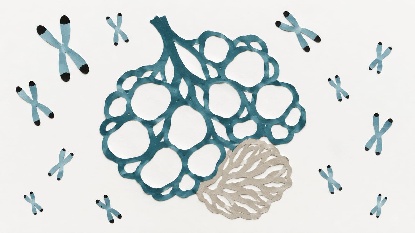
In telomere-length models, systems modeling aging through repeated cell division, and clinical biological samples, researchers tested a method for imaging individual telomeres. Telomeres are protective stretches of DNA at chromosome ends. Their lengths are measured in base pairs, DNA’s paired building blocks; a kilobase is 1,000 base pairs.
The method used a DNA-targeting tool to label telomere boundaries and magnetic beads to collect the DNA without copying it. An automated imaging workflow then measured individual telomeres rather than population averages. The assay used 500 nanograms of DNA and required 2–4 hours of hands-on processing. Measurements showed high reproducibility across repeated tests and different operators. The researchers also used DNA labels near telomeres to identify which chromosome arm they came from.
Why it matters
Critically short telomeres can trigger cellular stress and senescence, a state in which cells stop dividing. Measuring these unusually short chromosome ends may support the development of aging-related biomarkers.
Caveats
The abstract reports method validation, not evidence that these measurements predict aging or disease outcomes, and does not give validation sample sizes. This preprint has not been peer-reviewed.
The paper
Virginia Commonwealth University
Research Square, Oct 28, 2026, Open access, CC BY, Preprint, not peer-reviewed