Placentas showed faster molecular aging in late-onset preeclampsia
An analysis of placental tissue linked the pregnancy complication to shorter chromosome ends, DNA damage and cells that had stopped dividing.
PNAS
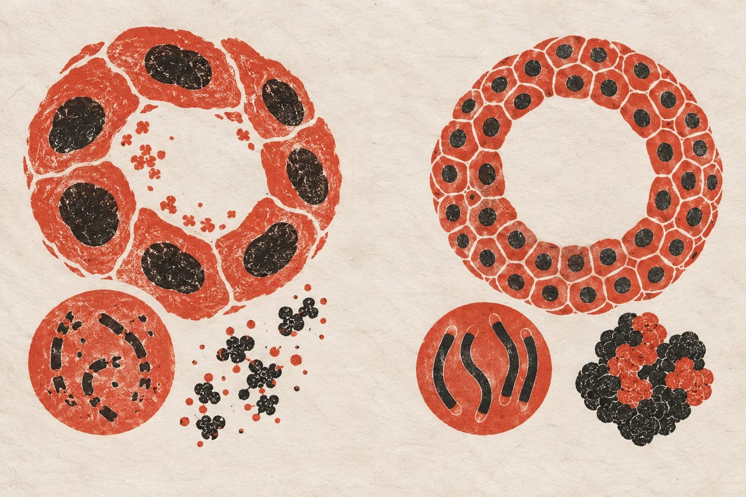
Researchers studied primary placental tissue and trophoblast organoids, lab-grown models made from cells that help form the placenta. They examined late-onset preeclampsia, a pregnancy complication involving high blood pressure and placental dysfunction. Affected placentas showed accelerated molecular aging, with DNA damage and shortened telomeres, the protective ends of chromosomes. Placental cells also showed senescence, a state in which cells stop dividing.
Experiments using tissue and organoids identified oxidative stress, stress caused by reactive oxygen molecules, as a driver of telomere shortening and an imbalance in blood-vessel growth signals. The antioxidant enzyme superoxide dismutase preserved telomere length, reduced DNA damage and restored this balance. Inflammation did not alter placental aging patterns. Levels of telomere-associated RNA molecules were also lower in affected placentas. Experimentally reducing these molecules worsened telomere erosion and senescence.
Why it matters
Placental decline in preeclampsia offers a setting for studying tissue aging. The work points to lab-grown placental models as a way to explore possible approaches to limiting preeclampsia-associated placental dysfunction.
Caveats
The study used placental tissue and laboratory models, not a trial in pregnant people. These results do not show whether antioxidants would safely or effectively treat preeclampsia.
The paper
Late-onset preeclampsia is characterized by accelerated placental aging
Show 7 more authors
Dulce L Medina Garcia, Ellen Menkhorst, German Mora, Jessica M Williamson, Lynda K Harris, Jose M Polo, David A MacIntyre,Flinders University
PNAS · 6 Oct 2026