Brain structure linked to memory-training gains in older adults
In 30 healthy adults aged 65–75, brain scans before four weeks of home-based memory training identified measures associated with training benefit.
4 weeks, non-randomized trial, independent replication
Limit: Only thirty older adults contributed to the prediction models
Edited by Boris Djordjevic · AgingResearch.News
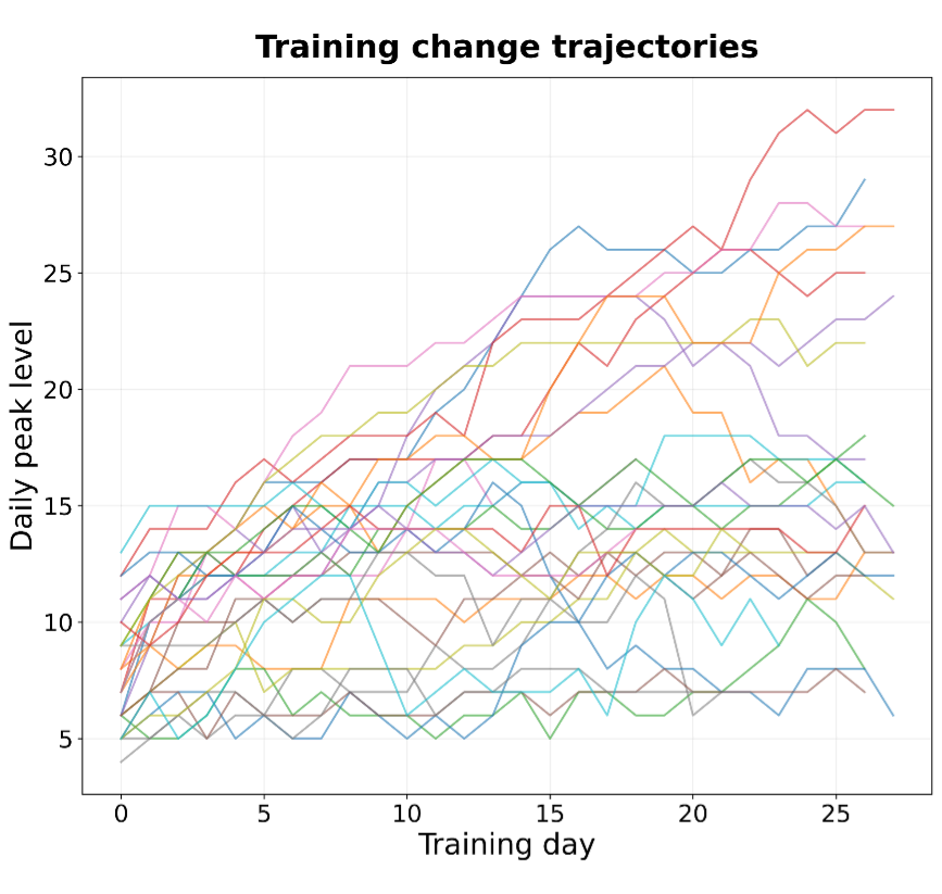
Thirty healthy adults aged 65–75 underwent magnetic resonance imaging (MRI) before four weeks of home-based memory training. The study analyzed associations between brain measurements and training gains. Participants practiced the Method of Loci, a memory technique that links information to imagined locations.
The volume of the left hippocampus, a brain region involved in memory, predicted training responsiveness; right hippocampal volume did not. Associations were concentrated toward the back of the hippocampus. An MRI measure of signals attributed to neurites, the extensions of nerve cells, also predicted benefit in the back but not the front. Signals attributed to nerve-cell bodies did not predict benefit. Both types of measures predicted responses when researchers left each participant out of the analysis in turn and tested the prediction on that person.
Why it matters
Memory for events often declines with age. Understanding differences in responsiveness to memory training may help inform personalized cognitive interventions for older adults.
Caveats
These associations do not show that brain structure caused differences in training benefit. The preprint has not been peer-reviewed and studied only 30 healthy adults aged 65–75.
- Method of loci training
- Right hippocampal volume
- Left hippocampal volume
- Neurite signal fraction
- Soma signal fraction
- Humans
The paper
University “G. d’Annunzio” of Chieti- Pescara
Research Square, Oct 20, 2026, Open access, Preprint, not peer-reviewed


