Brain scan model estimates aging across brain regions
Trained on scans from 14,423 adults with normal cognition, the model performed better than an existing method in repeat-scan testing.
Our read
Biomarkers- Studied:
- Computational study of 14,423 adults with normal cognition, against an existing method
- What changed:
- The brain-age model performed better on repeat scans than an existing method
- The catch:
- Brain-scan age estimates are stand-ins for biological aging, not direct measurements
Editor of record: Boris Djordjevic · AgingResearch.News
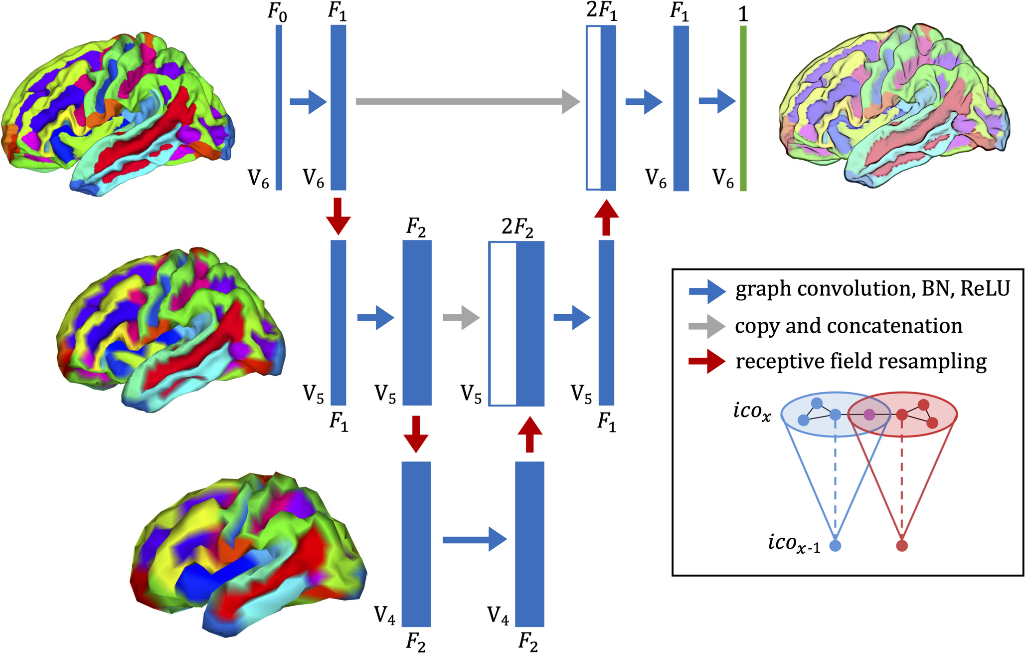
In a modeling study, researchers trained a computer model on brain scans from 14,423 adults with normal cognition. It used structural features of the cortex, the brain’s outer layer, to estimate brain age region by region. These features included thickness, surface area and curvature.
The model performed better than an existing method when tested on repeat scans. In people with mild cognitive impairment, it identified widespread estimated aging, particularly in a region called the parahippocampal gyrus. In people with Alzheimer’s disease, estimated aging extended across the entire cortex, particularly in its inner temporal regions. Differences between estimated local brain age and a person’s age were associated with measures of Alzheimer’s-related cognitive impairment.
Why it matters
A whole-brain age estimate summarizes anatomical aging in one number. Local estimates offer a way to investigate where age-related structural changes are concentrated within an individual brain.
Caveats
The findings concern model-based estimates from brain scans, not direct measurements of biological aging. The abstract does not report the sizes or age ranges of the groups with mild cognitive impairment or Alzheimer’s disease.
The paper
University of Southern California
Neuroinformatics, Oct 28, 2026, Open access

Comments
Be the first to comment.