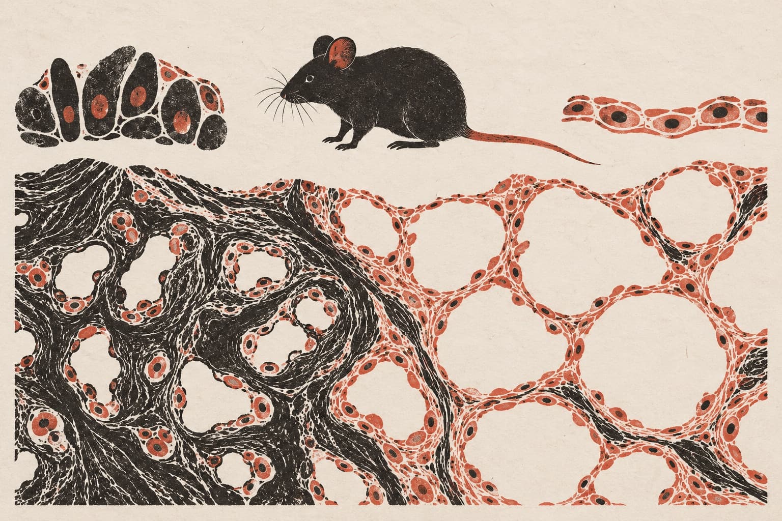
Cell changes may contribute to high lung blood pressure in rats
A four-week low-oxygen experiment in rats found changes in lung-lining cells and identified interventions that eased disease progression.
Limit: Lung-directed Smpd2 silencing does not isolate the contribution of air-sac cells
Edited by Boris Djordjevic · AgingResearch.News
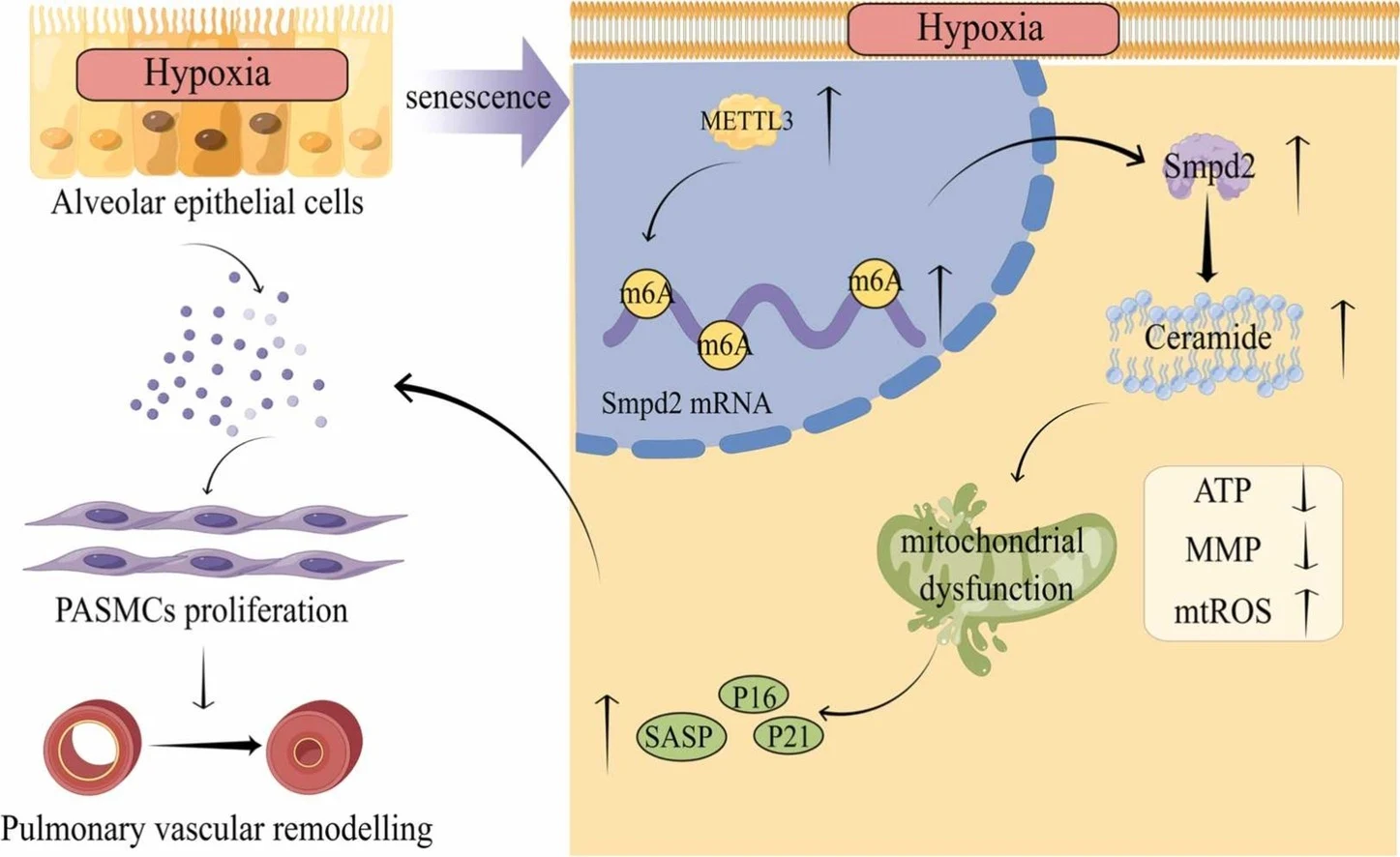
In rats, a four-week low-oxygen experiment examined high blood pressure in the lungs. Cells lining the lungs’ air sacs developed more pronounced signs associated with senescence, a state in which cells stop dividing. Reducing activity of the gene Smpd2 in the lungs reduced these changes and eased disease progression.
In human air-sac cells grown in the lab, low oxygen increased chemical marking of RNA from Smpd2, making the RNA more stable and increasing the gene’s activity. These changes were associated with buildup of ceramide, a fat molecule, and dysfunction of mitochondria, the cells’ energy-producing structures. Signals released by air-sac cells with senescence-associated changes were linked to abnormal growth of lung artery muscle cells in lab experiments. The findings suggested that these cell changes may contribute to high lung blood pressure.
Why it matters
The work connects research on cell senescence to disease in the lung’s blood vessels and points to candidate targets for future validation in humans.
Caveats
These were experiments in rats and human cells grown in the lab, not studies in people. The proposed mechanism needs validation in human disease and animal models targeting specific cell types.