Melatonin reduced senescence markers in human skin cells
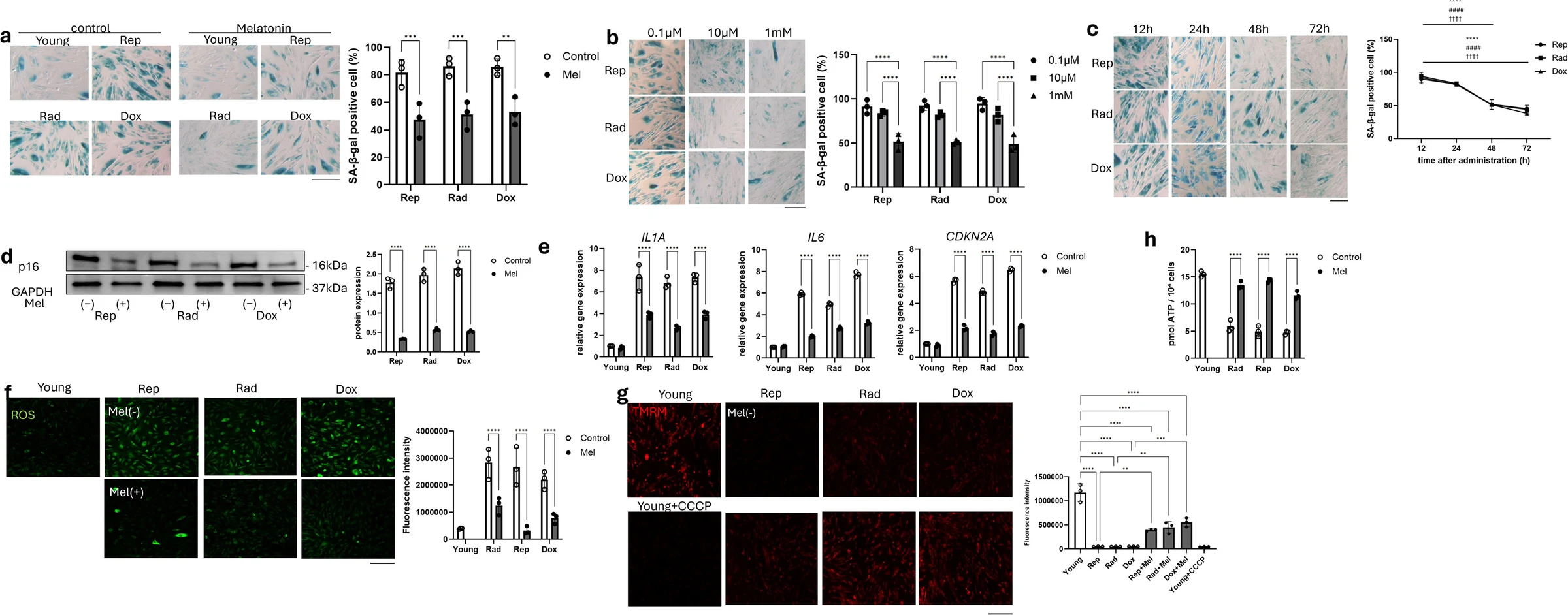
In human skin cell experiments, melatonin reduced the share of cells testing positive for a senescence marker by 38.5%–41.6%.
Limit: The abstract omitted doses, leaving the required melatonin exposure unspecified
In experiments with human dermal fibroblasts, cells that maintain the skin’s supporting structure, researchers tested melatonin in three models of senescence, a state in which cells stop dividing. They studied senescence caused by repeated cell division, irradiation or exposure to the drug doxorubicin.
Melatonin reduced the proportion of cells with a senescence marker. It also reduced inflammatory signals and markers of DNA damage, and restored measures of mitochondrial function. Mitochondria help supply cells with energy. In the human cells, melatonin lowered CXCL9, an inflammation-related signaling protein. Reducing production of this protein mimicked melatonin’s effects on senescence. In aged mouse skin, melatonin injections increased the thickness of the skin’s inner layer by 97% and the density of collagen, a structural protein, by 23%.
Why it matters
Fibroblast senescence contributes to the breakdown of the skin’s supporting structure with age. Understanding its links to inflammation and cellular energy supply may help guide research on skin aging.
Caveats
These were laboratory experiments in human cells and aged mouse skin, not a study of melatonin use in people. The abstract did not report doses, treatment durations or sample sizes.
Melatonin is part of Rhonda Patrick's supplement routineMore supplements
The paper
Keio University School of Medicine; Long School of Medicine
The Journal of Investigative Dermatology, Oct 19, 2026


