Chest X-rays help flag very low bone density in the lower spine
Among 3,007 adults, artificial intelligence identified 91.0% of those with very low lower-spine bone density and correctly classified 82.7% of those without it.
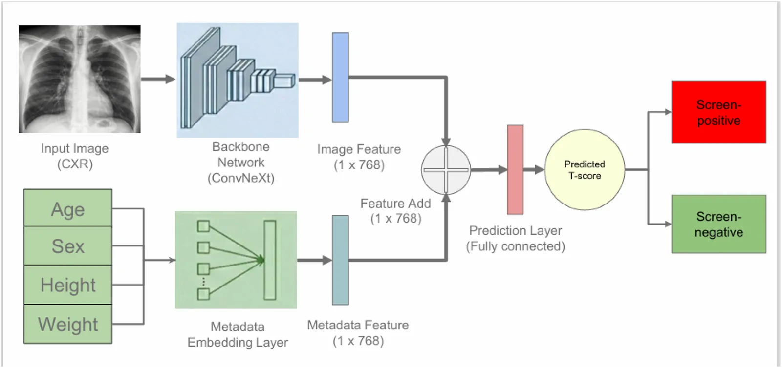
An observational validation study tested an artificial intelligence system in 3,007 adults who had a chest X-ray and a bone-density scan on the same day at two institutions. Researchers trained it on 5,943 paired examinations without collecting raw images centrally.
Bone scans identified very low bone density in the lower spine in 200 participants (6.7%). The system scored 0.942 on a measure of how well it distinguished adults with and without this finding. It identified 91.0% of those with very low density and correctly classified 82.7% of those without it. Performance held across sites, sexes and adults aged 60 or older. A version using images alone performed better than one using clinical information alone, supporting an imaging contribution beyond demographics.
Why it matters
Osteoporosis is an age-related bone disease. Using images already collected during routine care may offer a way to widen screening without centralising raw patient images.
Caveats
This observational validation assessed a scan-defined finding, not whether screening improved health. The authors positioned the system as an aid to selecting people for confirmatory bone-density scans, not a replacement for those scans.
- Deep learning
- Osteoporotic-level bone mineral density
- Lumbar spine bone mineral density
- Lumbar spine DXA T-score
- Lumbar-spine DXA-defined osteoporotic-level bone mineral density
- Humans
The paper
National Taiwan University; E-Da Hospital
Osteoporosis and Sarcopenia, 26 Jun 2026, Open access