Computer model detected fractures and located bones in spine scans
The model scored 0.952 for distinguishing scans with fractures from those without, compared with 0.913 for an earlier detection-only model.
Osteoporosis and Sarcopenia
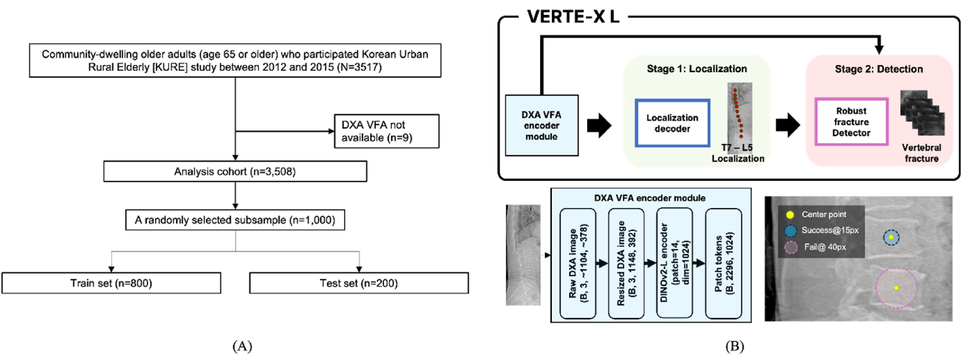
In a computational study, researchers used spine scans from 1,000 community-dwelling adults aged 65 or older in the Korean Urban Rural Elderly cohort. The images came from dual-energy X-ray absorptiometry, a type of bone scan used here to assess spine fractures. They trained the model on scans from 800 participants and tested it on scans from the remaining 200.
The model both located individual spine bones and detected fractures. Its score for distinguishing scans with fractures from those without was 0.952, compared with 0.913 for an earlier detection-only model. It identified 89.7% of scans with fractures and correctly classified 94.2% of scans without them. It located 94.7% of fractured spine bones within 15 pixels of their reference position.
Why it matters
The work focused on assessing osteoporotic spine fractures in older adults. It bears on how computer models might support that assessment.
Caveats
This computational study measured performance on images, not benefits for patients. The test set contained 200 participants from one Korean cohort.
The paper
Yonsei University
Osteoporosis and Sarcopenia · 1 Sep 2026 · CC BY-NC-ND