Plant-Derived Senolytic Ingredient, Camellia sinensis Root Extract, Ameliorates Skin Aging-Associated Phenotypes
Abstract
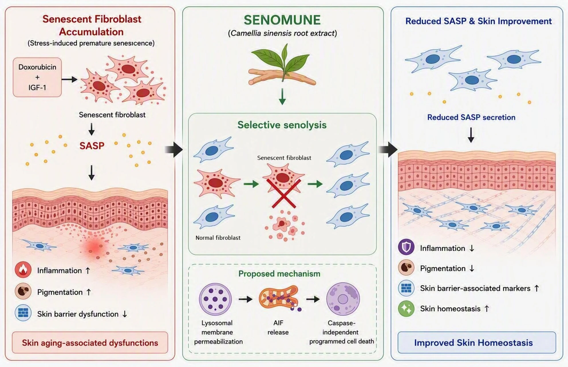
Senescent cells affect neighbouring cells via the senescence-associated secretory phenotype (SASP), which involves proinflammatory cytokines, chemokines and proteases. Camellia sinensis root extract (SENOMUNE) has shown therapeutic potential for skin aging-related disorders. However, whether it exerts senolytic effect remains to be explored. Thus, this study aimed to assess the senolytic properties of SENOMUNE in a stress-induced premature senescence model in normal human dermal fibroblasts (NHDFs) through cell viability assays, quantitative reverse transcription polymerase chain reaction, western blot and flow cytometry. NHDFs were induced to senescence using doxorubicin and insulin-like growth factor-1. The senolytic effect of SENOMUNE was evaluated by measuring senescence-associated β-galactosidase activity, gene and protein expression, and the apoptosis rate. SENOMUNE reduced the viability of senescent cells in a dose-dependent manner, with a more pronounced effect in senescent cells than in control cells. It was also associated with caspase-independent programmed cell death involving apoptosis-inducing factor and lysosomal membrane permeabilization in senescent cells. Moreover, SENOMUNE reduced the production of SASP factors, increased the expression of skin barrier-associated markers, and attenuated pigmentation by modulating the secretion of inflammatory cytokines. These effects were evaluated using conditioned media derived from senescent fibroblasts and validated through ex vivo skin biopsy experiments. Our results suggest that SENOMUNE exhibits senolytic properties and thus is a promising anti-aging phytopharmaceutical ingredient for managing skin-related disorders.
From the paper
- Result
… as evidenced by a leftward shift of the dose–response curve and a reduction in the estimated IC₅₀ from approximately 161 ppm (95% CI, 134–188 ppm) in control cells to 129 ppm (95% CI, 101–157 ppm) in DI cells (Figure S2).
Quoted word for word from the full text on onlinelibrary.wiley.com.
The paper
Research and Innovation Center
Experimental Dermatology, 1 Oct 2026