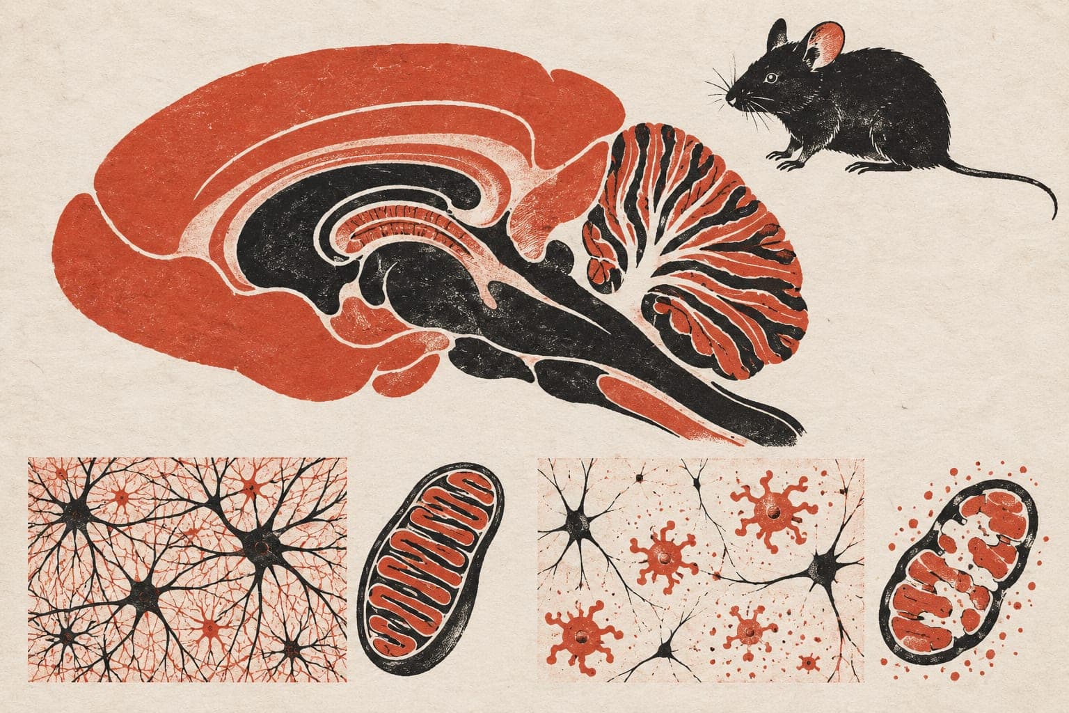
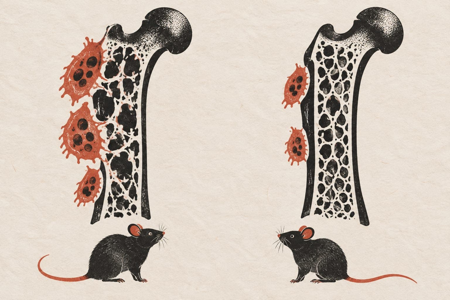
Deleting a signaling protein limits bone loss in mice
In mice lacking ovaries to mimic estrogen loss, deleting the Camkk1 gene from bone-degrading cells attenuated the loss of spongy bone.
Pharmacological Research
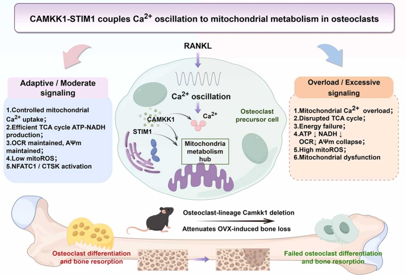
In an experiment using cultured cells and mice, researchers investigated how bone-resorbing cells called osteoclasts respond to calcium signals. In lab-grown osteoclasts, reducing levels of the protein CAMKK1 diminished calcium fluctuations, calcium uptake by energy-generating mitochondria, and osteoclast formation, while boosting CAMKK1 had the opposite effect. The team identified STIM1, another calcium-regulating protein, as a CAMKK1-associated partner. Depleting STIM1 also attenuated calcium signals, mitochondrial respiration, and osteoclast differentiation. When researchers forced cells to overexpress both proteins, this caused mitochondrial calcium accumulation, oxidative stress, membrane-potential disruption, respiratory dysfunction, and cell death. Finally, in mice that had their ovaries removed to model estrogen deficiency, deleting Camkk1 in the osteoclast lineage attenuated the loss of spongy trabecular bone.
Why it matters
Bone loss accelerates after menopause as estrogen levels decline, driven in part by overactive osteoclasts. Understanding how these cells coordinate calcium signals and energy metabolism bears on how bone deteriorates during estrogen deficiency.
Caveats
Much of the signaling mechanism was evaluated in cultured cells, and the living-animal findings rely on surgically induced hormone loss in mice. Whether targeting this pathway safely alters bone density in humans remains untested.
The paper
Show 4 more authors
Yuan Liu, Xuefang Zhang, Shuaijun Jia, Liang Yan,Honghui Hospital
Pharmacological Research · 8 Oct 2026