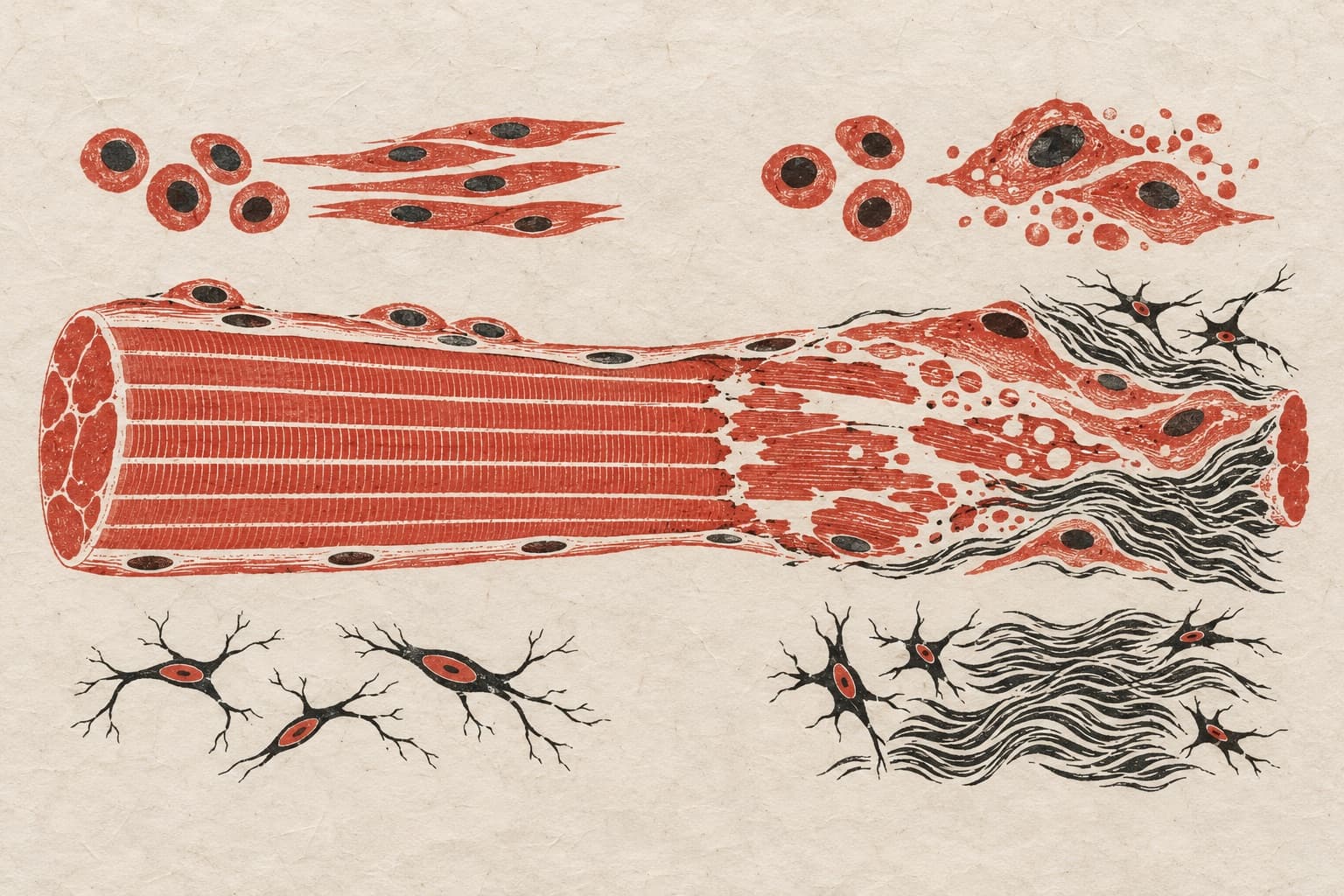
Human lymph node fibroblasts show cellular aging features before rheumatoid arthritis onset
Fibroblasts from individuals at risk for rheumatoid arthritis exhibit impaired adipogenic differentiation and downregulated DNA repair and cell cycle pathways.
RMD Open
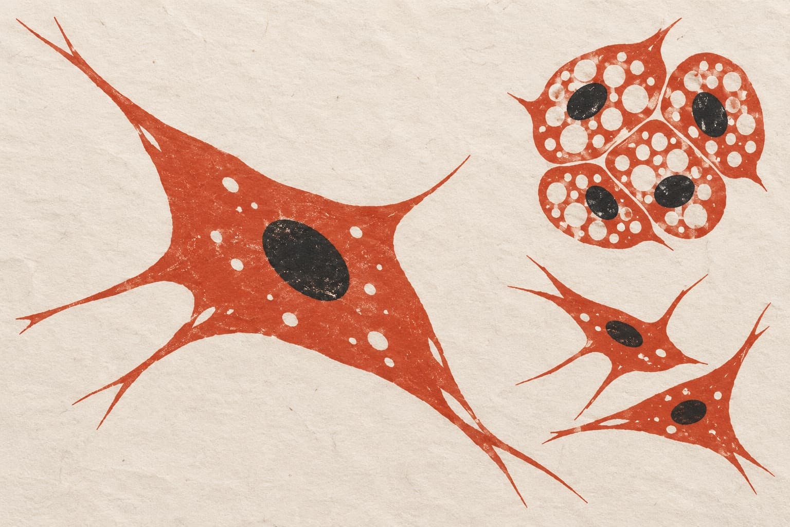
In a human cell study, researchers analyzed primary lymph node fibroblasts from healthy controls, individuals at risk of rheumatoid arthritis, and patients with established disease. The team isolated cells from inguinal lymph node core biopsies to evaluate differentiation capacity and age-associated alterations. Using adipogenic differentiation assays, they tested cellular plasticity, while bulk RNA sequencing tracked transcriptional shifts. Fibroblasts from at-risk individuals and rheumatoid arthritis patients showed impaired adipogenic capacity, forming fewer lipid droplet-positive cells upon differentiation. Transcriptomic analyses revealed the downregulation of cell cycle, DNA repair, and differentiation pathways. In addition, the cells failed to upregulate the cell cycle switch G0S2 upon stimulation. These coordinated defects indicate that features of cellular aging emerge prior to clinical rheumatoid arthritis onset.
Why it matters
The findings indicate that cellular aging signatures in structural lymph node cells precede systemic inflammatory symptoms. This suggests that early declines in stromal cell fitness could contribute to the loss of immune homeostasis in age-associated autoimmune diseases.
Caveats
The findings rely on in vitro functional assays and bulk RNA sequencing from biopsy-derived cells rather than in vivo tissue models. The abstract also does not provide sample sizes or report longitudinal outcomes showing which at-risk individuals ultimately developed rheumatoid arthritis.
- DNA repair
- Cell cycle regulation
- Stem cell differentiation
- Rheumatoid arthritis
- Lipid droplet-positive cells
- Humans
The paper
Amsterdam UMC Location University of Amsterdam
RMD Open · 1 Oct 2026