ADA loss impairs DNA repair and spurs senescence in astrocytes
Studying sporadic ALS astrocyte models, researchers found TDP43 dysfunction impaired adenosine deaminase, disrupting purines and raising P16 levels while blunting 53BP1-mediated repair.
International Journal of Molecular Sciences
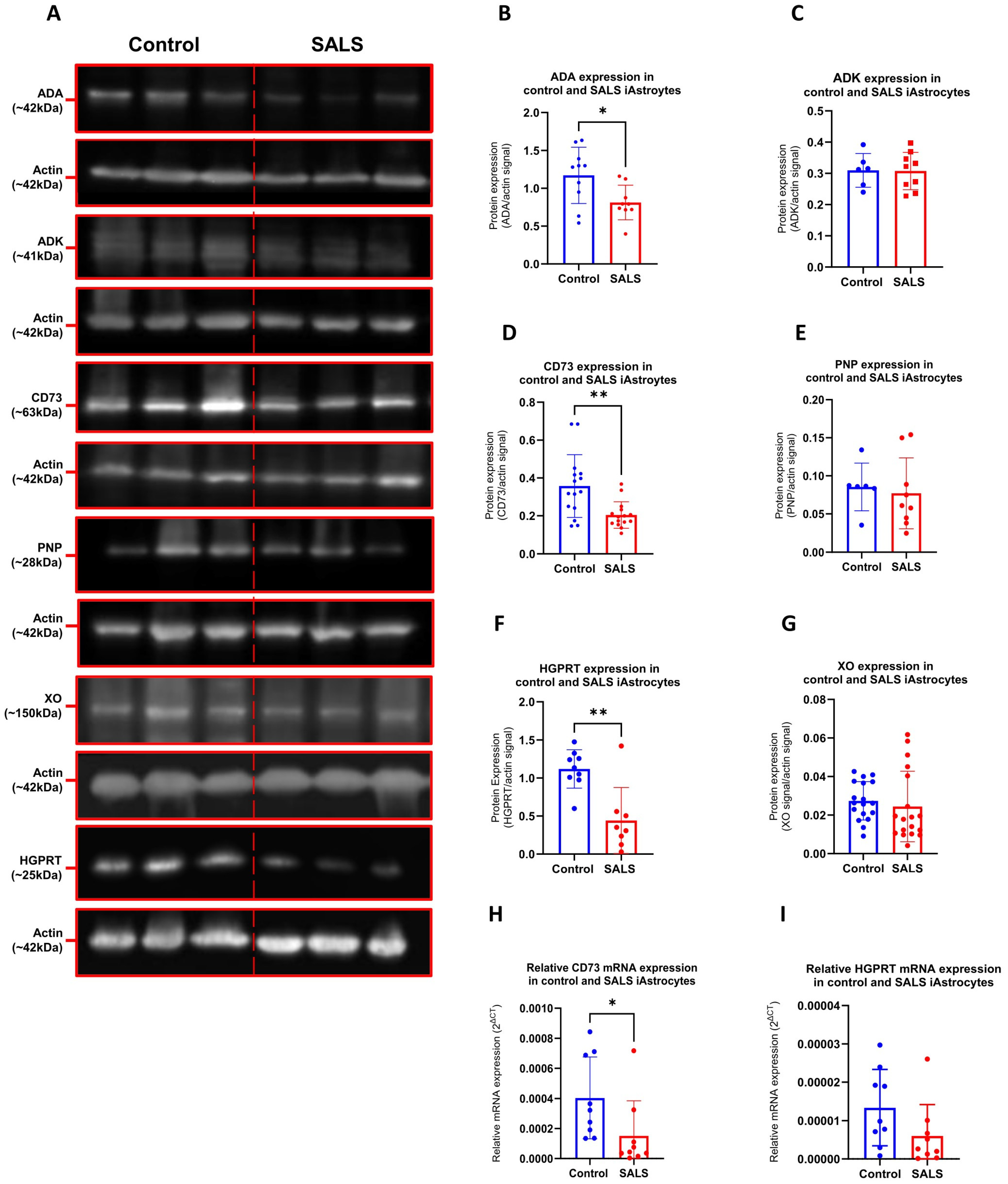
In human cell models and patient tissues, researchers investigated purine metabolism dysfunction in sporadic amyotrophic lateral sclerosis (ALS). In induced neural progenitor cell-derived astrocytes from sporadic ALS cases, loss of the enzyme adenosine deaminase (ADA) disrupted purine metabolite balances alongside alterations in ecto-5'-nucleotidase and hypoxanthine-guanine phosphoribosyl transferase. Loss or pharmacological inhibition of ADA reduced 53BP1-mediated DNA repair and increased levels of the senescence marker P16. In vitro, TDP43 dysfunction drove this purine metabolism impairment, yielding DNA damage—likely through the inhibition of repair pathways—and cellular senescence. Disrupted purine profiles were also detected in patient cerebrospinal fluid and post-mortem tissue, where downstream metabolite levels varied with age and sex and positively correlated with disease progression.
Why it matters
The results link purine metabolic defects to impaired DNA repair and glial senescence in ALS pathology. Targeting the ADA pathway may offer a candidate approach for slowing disease progression in neurodegenerative disorders.
Caveats
Mechanistic conclusions depend on in vitro cell cultures, and patient fluids and post-mortem tissues provide observational associations without longitudinal tracking. The abstract also does not report sample sizes or the magnitude of metabolite changes and senescence induction.
The paper
Show 19 more authors
Sangeet Makhija, Brittany C. S. Ellis, Kari E. Wong, Heather J. Walker, Miriam Yagüe-Capilla, Hannah O. Timmons, Arian Bradley, Ella Nightingale, Rees Ross, Chloe F. Allen, Noemi Gatto, Herbie Garland, Nikita Soni, Stephen J. Kolb, J. Robin Highley, Guillaume M. Hautbergue, Sean G. Rudd, Ryan J. H. West, Pamela J. Shaw,University of Sheffield
International Journal of Molecular Sciences · 16 Sep 2026 · CC BY