Software detected esophageal cancer on existing chest scans
In external testing of 11,466 patients, the model detected 90.0% of cancers and 52.5% of precancerous lesions, while correctly classifying 98.5% without either.
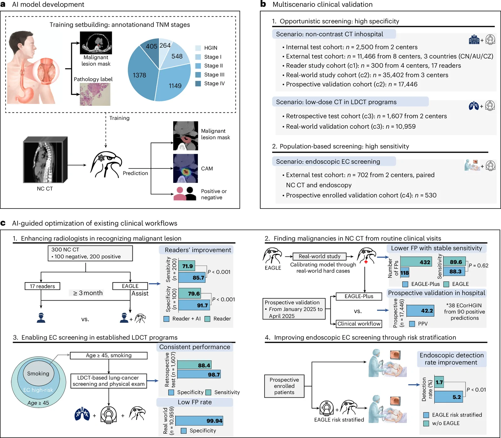
80,612 patients, multicenter, registered ChiCTR2300074806
Limit: Follow-up in the prospective and real-world validations was less than 2 years
Edited by Boris Djordjevic · AgingResearch.News
In an observational study, researchers trained an artificial intelligence model on chest scans from 6,813 patients. They validated it in 80,612 patients across 12 centers in three countries. The model analyzed computed tomography scans—detailed X-ray images—taken without contrast dye. It looked for cancer and precancerous lesions in the esophagus, the tube carrying food to the stomach.
In external testing involving 11,466 patients, it detected 90.0% of cancers and 52.5% of precancerous lesions. It correctly classified 98.5% of patients without either condition. It detected 60.1% of early-stage cancers classified as stage I in that group. In a separate prospective hospital validation involving 17,446 patients, 42.2% of positive results were confirmed as cancer or precancer.
Why it matters
One screening cohort included adults aged 45–75, making the work relevant to cancer detection in midlife and later life. The authors suggest existing scans could support scalable early esophageal cancer screening.
Caveats
This observational validation does not establish whether screening improves survival. Some patients were classified as negative through clinical follow-up, which the authors acknowledged could have missed slow-growing lesions.
From the paper
- Participants
The RW1 cohort enrolled 20,758 consecutive patients from three centers, of whom 285 were diagnosed with esophageal malignancies.
Results, Model calibration and real-world retrospective validation
- Result
In the internal test set of 515 NC CT scans with lesion annotations (Extended Data Fig. 1), the mean lesion segmentation of Dice similarity coefficient (DSC) was 0.744 (95% CI = 0.722–0.766).
- Limitation
First, the diversity of the training and validation populations requires further expansion.
Quoted word for word from the full text on nature.com.
The paper
Sun Yat-sen University; Sichuan Cancer Hospital
Nature Medicine, Sep 22, 2026, Open access


Comments
Be the first to comment.