Strength workouts linked to different brain changes in older men
A 12-week randomized trial in 63 older men linked heavier lifting and blood-flow-restricted exercise to changes in different brain-scan measures.
63 older men
Limit: Excluding participants without post-intervention MRI scans may have introduced healthy completer bias
Edited by Boris Djordjevic · AgingResearch.News
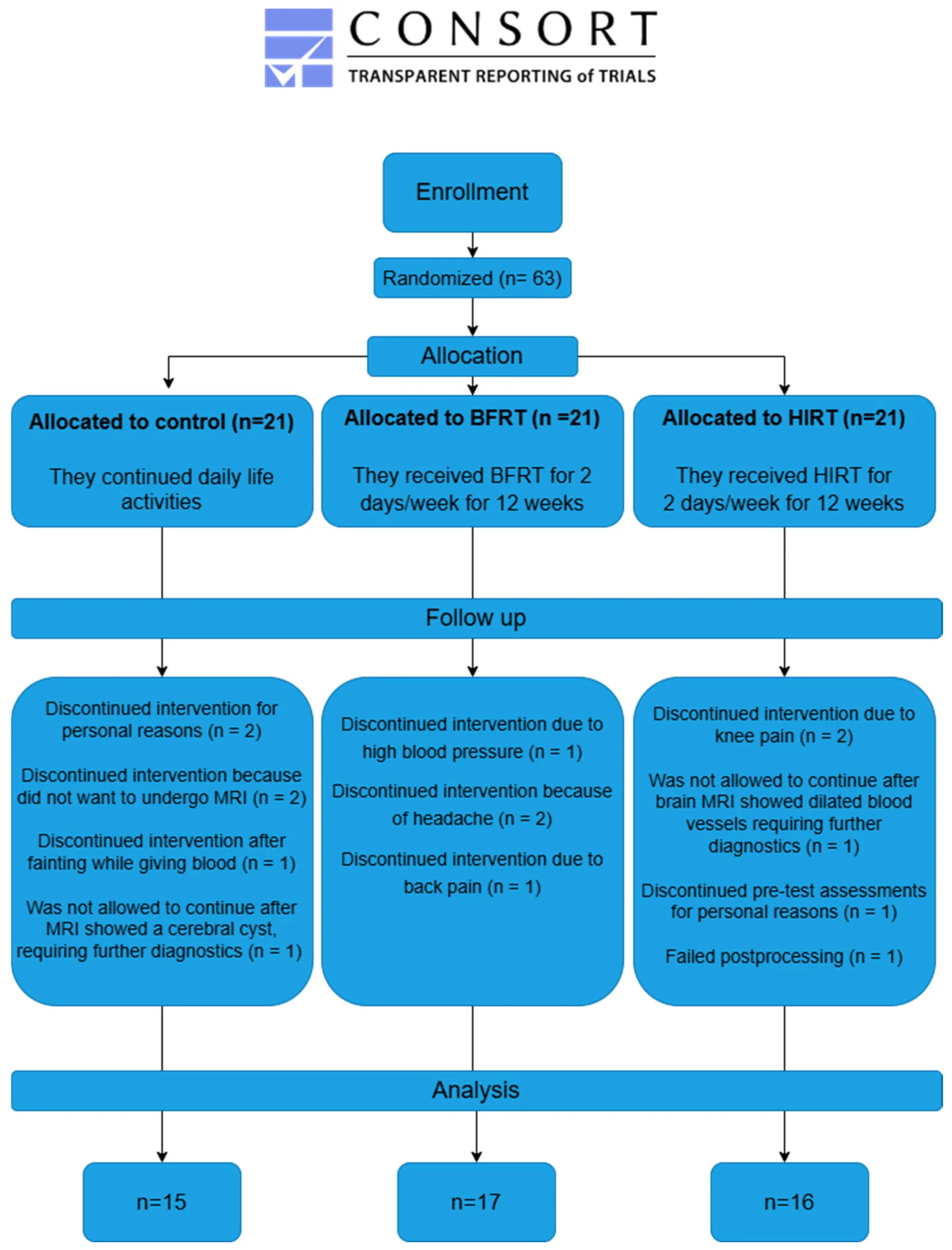
An exploratory randomized trial assigned 63 community-dwelling men aged 60–78 to two strength-training programs or a control group for 12 weeks. Both programs involved three leg exercises twice weekly. Cuff-assisted training restricted blood flow while men lifted 40% of their maximum load. High-intensity training combined lifting at 80% with lowering at 120%, with coaches raising the heavier weights.
Among 48 men with before-and-after brain scans, high-intensity training was associated with thickness changes in the brain’s outer layer in three left-sided regions compared with controls. Cuff training was associated with changes in a scan measure of nerve-fiber organization in three regions. These regions were in the parietal area, toward the brain’s top and back. Training-related withdrawals involved increased blood pressure (one man), headaches (two) and back pain (one) with cuffs, and knee pain (two) with high-intensity training.
Why it matters
The study addressed whether resistance exercise can influence brain structure in later life, beyond its effects on muscle.
Caveats
This exploratory pilot analyzed only men who completed both scans; structural scan changes do not establish better thinking or slower brain aging. The findings may not apply to women or people with excluded conditions, including cognitive, neurological or cardiovascular disorders and diabetes.
From the paper
- Participants
A total of 63 older men (age range: 60–78) with a BMI ranging between 21.3 kg/m² and 32.9 kg/m² and handgrip strength ranging between 34 and 60 kg were included.
- Who took part
The BRAIN-M trial, conducted at the Lithuanian Sports University in Kaunas, Lithuania, enrolled sixty-three community-dwelling males aged more than 60 years between October 2022 and November 2023.
- Duration
The subjects in the intervention groups exercised twice a week for twelve weeks.
Methods, 2.6. Intervention: Resistance Exercise Intervention
- Limitation
This study was designed as an exploratory pilot study; therefore, no formal a priori sample size calculation was performed.
- Cortical thickness
The parietal cortical regions demonstrating these changes were the left inferior parietal cortex (p = 0.049), left posterior cingulate cortex (p = 0.028) and left supramarginal cortex (p = 0.018); for illustration, see.
- Fractional anisotropy
Post hoc analysis revealed significant differences between the BFRT and control groups in the following parietal regions: left inferior parietal (p = 0.040), left precuneus (p = 0.040) and right superior parietal (p = 0.011).
Quoted word for word from the full text on PubMed Central.
- Blood flow restriction training
- High-intensity resistance training
- Cortical thickness
- Fractional anisotropy
- Thigh muscle anatomical cross-sectional area
- Humans
The paper
Lithuanian University of Health Sciences; Lithuanian Sports University
Brain Sciences, Jul 28, 2026, Open access


Comments
Be the first to comment.