Imaging the neurohiv brain in animal models: past, present, and future-toward longitudinal, whole-brain aging phenotypes
Abstract
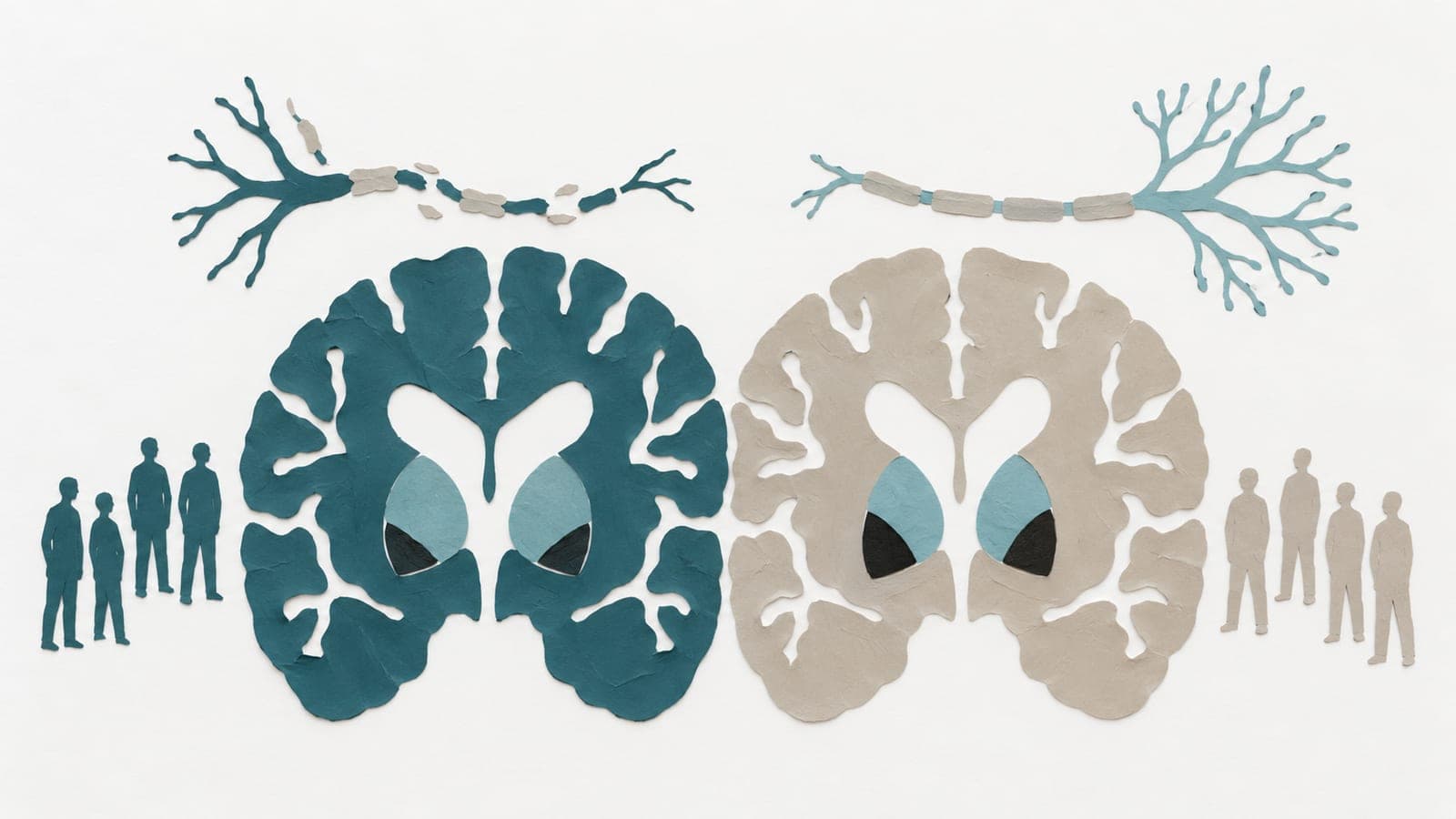
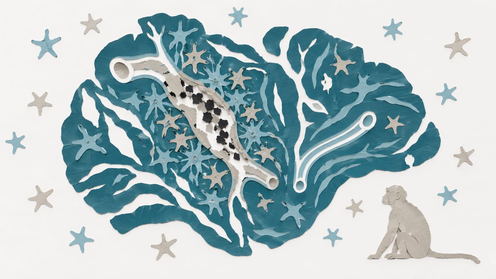
Animal models are essential for defining mechanisms of HIV-associated brain injury in the antiretroviral therapy era, where neurocognitive impairment and brain aging have replaced opportunistic infections as dominant clinical concerns. Humanized mice, EcoHIV murine systems, and SIV or SHIV nonhuman primates now enable increasingly sophisticated brain imaging across in vivo and ex vivo scales. Structural and diffusion MRI, perfusion imaging, manganese-enhanced MRI, and multiphoton and endoscopic optical methods provide complementary sensitivity to tissue integrity, hemodynamics, cellular dynamics, and neurovascular coupling. Ex vivo clearing and light-sheet microscopy further support whole-brain mapping and atlas-based integration with histology. We synthesize how neuroHIV imaging has progressed from structural surrogates to quantitative, multimodal pipelines and highlight a central gap for neurocognitive aging research. Longitudinal diffusion and perfusion MRI in SIV-infected macaques demonstrate that within-animal imaging can capture infection-associated trajectories. In contrast, systematic in vivo whole-brain neuroimaging remains limited in widely used murine neuroHIV models, particularly EcoHIV, despite strong behavioral and virologic phenotyping. We argue that the next phase of the field requires standardized, longitudinal, multiscale pipelines that couple high-field MRI and targeted molecular imaging with mesoscopic optical and endoscopic approaches and whole-brain three-dimensional mapping frameworks. Such integration should enable imaging-derived brain aging phenotypes in neuroHIV models, clarify mechanisms linking persistent inflammation to neurovascular and circuit dysfunction, and accelerate intervention-ready biomarkers aligned with aging cohorts of people with HIV.
The paper
Pennsylvania State University
Frontiers in Aging Neuroscience, 1 Jul 2026, Open access