Tobacco smoke makes mouse lung cells stop dividing
Experiments in mice linked smoke exposure to disrupted cell clocks and immune activation, alongside early changes that may leave lungs prone to scarring.

Our read
Mechanisms- Studied:
- Experiments in mice exposed to environmental tobacco smoke
- What changed:
- Mouse lung cells stopped dividing after environmental tobacco smoke exposure
- The catch:
- Direct measurements of lung-cell division are not reported
- In the record:
- The 1,989th paper on smoking with results in people; 4,845 on record in all.
Editor of record: Boris Djordjevic · AgingResearch.News
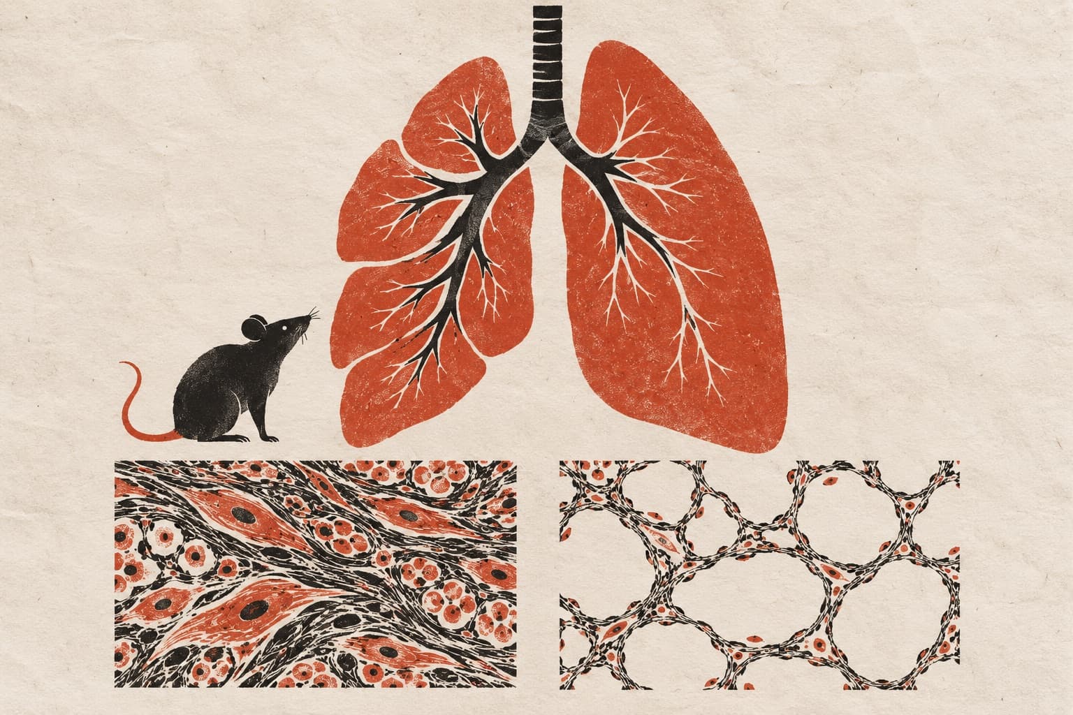
Mouse lungs exposed to environmental tobacco smoke developed senescence, a state in which cells stop dividing, in experiments examining links between cell clocks and lung injury. Smoke induced senescence in surface-lining cells and connective-tissue cells, even when the molecular clock remained intact. These cells also produced substances associated with senescence.
The researchers identified a shared gene-activity pattern across several lung cell types. It combined suppressed clock activity, senescence-related changes and activation of a pathway that senses DNA and triggers immune responses. Mice lacking two clock regulators also showed changes in these programs. Separately, mapping gene activity across smokers’ lungs revealed parallel changes, particularly in certain lung-lining cells and connective-tissue cells. The authors interpreted the findings as early tissue remodeling and preparation for scarring.
Why it matters
Senescence and DNA damage contribute to smoke-related lung injury. The authors interpret the combined program as an early checkpoint for susceptibility to lung scarring.
Caveats
The human gene-activity maps leave open whether clock disruption preceded senescence or followed it. The experiments testing these processes used mice.
The paper
University of Rochester Medical Center
International Journal of Molecular Sciences, Nov 15, 2026, CC BY

Comments
Be the first to comment.