Age links to brain fiber size differ among crossing fibers
Brain scans from 295 older adults linked age most strongly to smaller bundle size in the non-dominant groups of crossing nerve fibers.
Our read
Biomarkers- Studied:
- Cross-sectional study of 295 older adults
- What changed:
- Brain fiber-bundle size was lower at older ages, with steeper age associations in non-dominant than primary fibers
- The catch:
- Crossing-fiber complexity was confounded with tract identity and location
- In the record:
- One of 3 papers on fiber-bundle cross-section on record: 2 with results in people.
Editor of record: Boris Djordjevic · AgingResearch.News
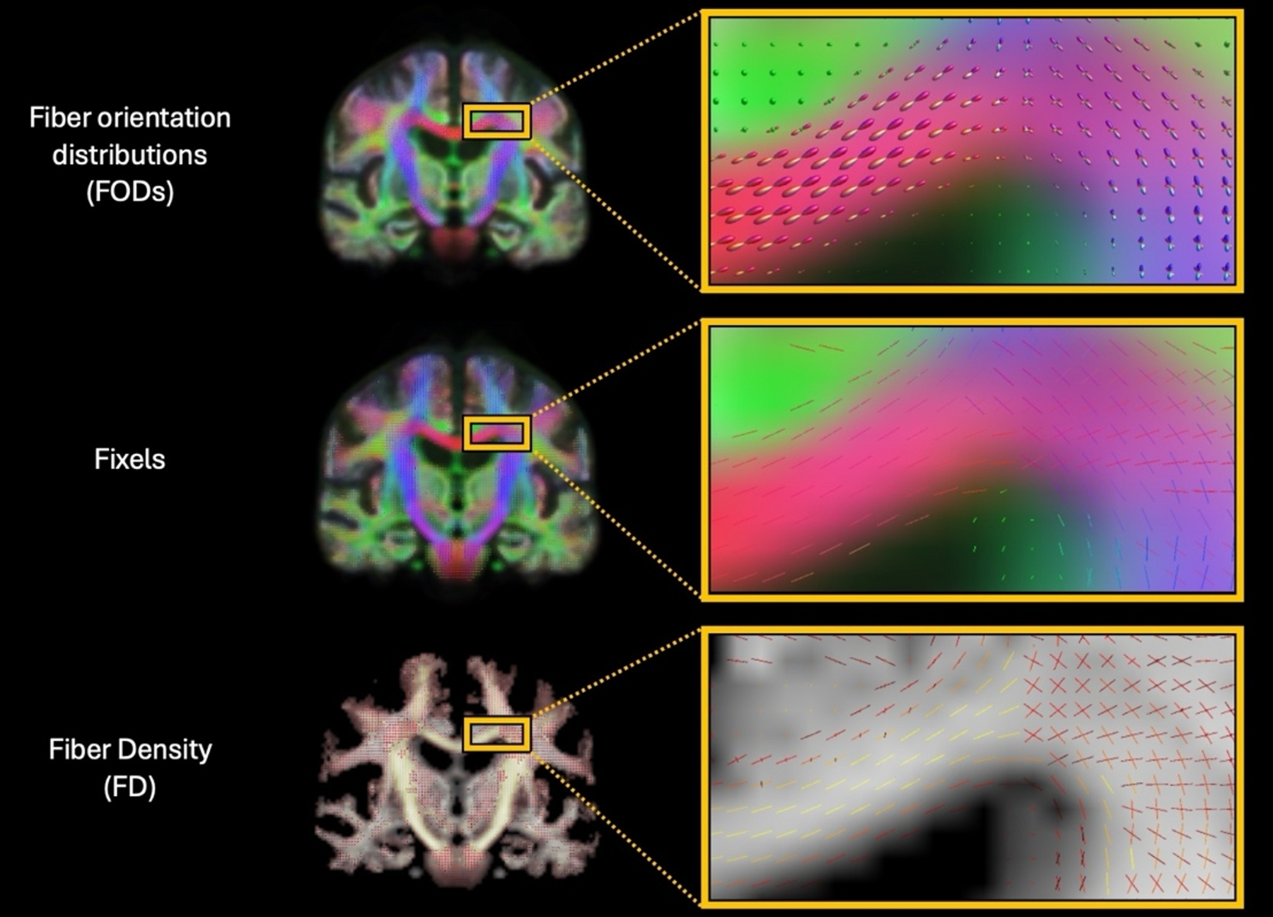
Among 295 adults aged 60 or older, a cross-sectional brain-scan analysis examined bundles of nerve fibers. Participants in the longitudinal observational Vanderbilt Memory and Aging Project were cognitively unimpaired or had mild cognitive impairment, meaning thinking or memory difficulties beyond normal aging. The analysis separated primary, secondary and tertiary fiber populations where fibers crossed. It measured bundle cross-section, a measure of bundle size.
Older age was linked to smaller cross-sections, most strongly in the secondary and tertiary, or non-dominant, populations. In these populations, the age links did not vary with the number of crossing bundles. Cross-sections were also smaller in participants with mild cognitive impairment than in unimpaired participants. Larger cross-sections in these populations were associated with better executive function—the mental skills used to plan and manage tasks.
Why it matters
The authors’ framework could help researchers study white matter—the brain tissue containing nerve fibers—in aging using existing brain-scan cohorts.
Caveats
Fiber measurements came from a single baseline scan per person, so the age associations did not measure changes within individuals. Participants were 60 or older and did not have dementia.
From the paper
- Participants
The analytic sample comprised 295 older adults from VMAP.
- Result
For the executive function composite, FC was positively associated with performance in N2 (β = 0.53, 95% CI [0.22, 0.84], t = 3.35, pFDR = 0.008) and N3 (β = 0.57, 95% CI [0.21, 0.93], t = 3.10, pFDR = 0.010).
Quoted word for word from the full text on link.springer.com.
- Mild cognitive impairment
- Executive function
- Fiber density
- Fiber density–cross-section composite
- Fiber-bundle cross-section
- Humans
The paper
Brain Structure & Function, Nov 14, 2026, Open access, CC BY
Comments
Be the first to comment.