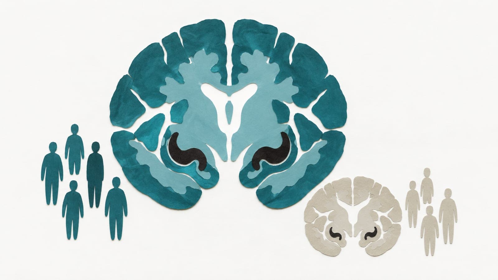
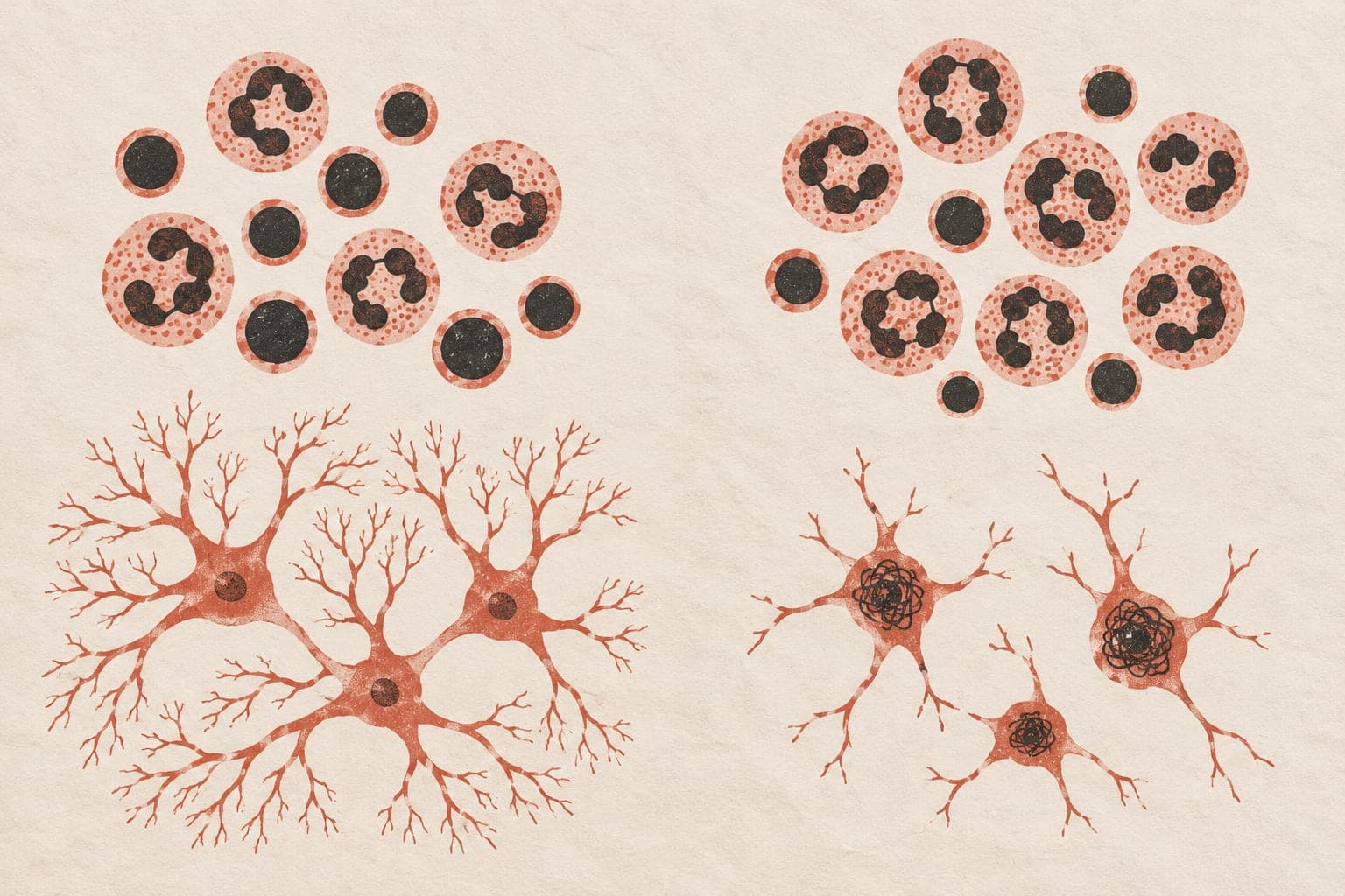
Brain MRI score fell across Alzheimer’s disease stages
The score distinguished stable normal cognition from prediagnosis Alzheimer’s with an area under the curve—a measure of group separation—of 0.817.
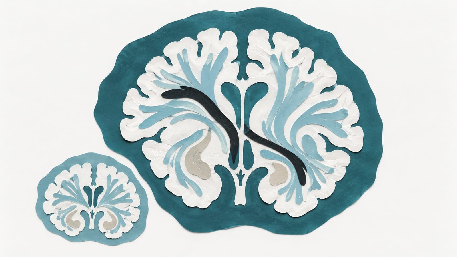
1,706 participants, independent replication
Limit: The combined model’s positive predictions were only modestly reliable
Edited by Boris Djordjevic · AgingResearch.News
An observational study analyzed 1,706 brain-scanning sessions from the OASIS-3 dataset. Researchers used diffusion MRI, which maps water movement, to assess routes through white matter, the nerve fibers connecting brain regions. Their Route Concordance Index was lower before clinical conversion and declined steadily across five disease stages.
The score distinguished people who remained cognitively normal from those assessed before Alzheimer’s diagnosis about as well as hippocampal volume, the size of a memory-related brain region. Findings replicated in the independent ADNI dataset. A model combining routing measures with demographic information, brain-structure measurements and cognitive scores identified conversion to mild cognitive impairment or Alzheimer’s disease up to four years before diagnosis.
Why it matters
Alzheimer’s pathology accumulates long before diagnosis. Measuring changes in white matter may help address how far the disease has advanced, rather than simply whether that pathology is present.
Caveats
This observational analysis needs validation in studies that follow people forward before clinical use. The preprint has not been peer-reviewed, and the combined model’s positive predictions were only modestly reliable.
- Machine learning
- Alzheimer's disease
- Route Concordance Index
- Hippocampal volume
- Conversion to mild cognitive impairment or Alzheimer's disease
- Humans
The paper
University of California san Diego
medRxiv, Oct 24, 2026, Open access, CC BY, Preprint, not peer-reviewed