Sugar patterns on proteins varied with age in mouse tissues
An atlas of 80,618 sugar-bearing protein fragments covered nine ages across the mouse lifespan and revealed changes at specific attachment sites.
Limit: Sugar patterns between the nine sampled ages were not measured
Edited by Boris Djordjevic · AgingResearch.News
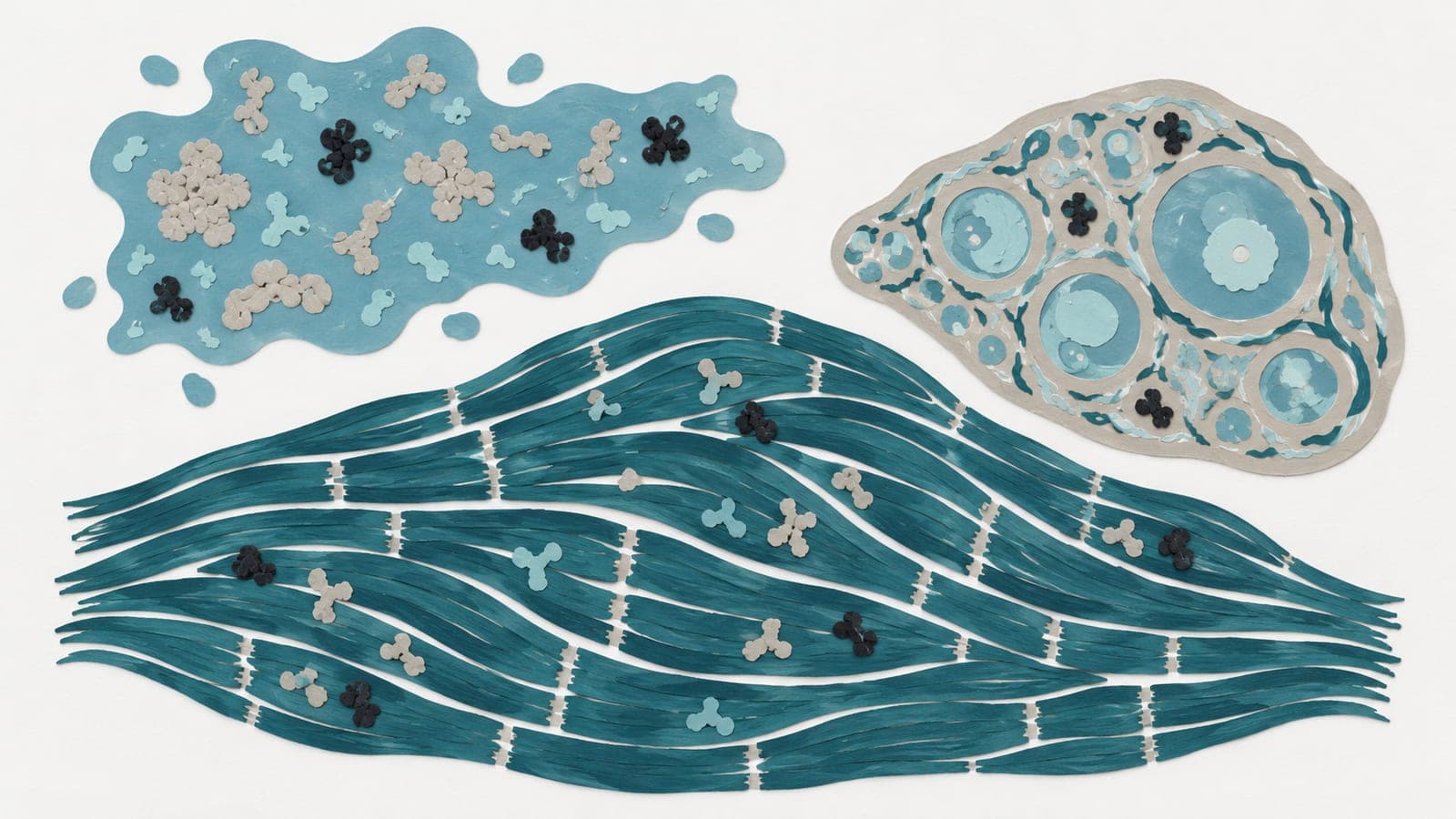
In a laboratory mapping study, researchers examined sugars attached to proteins in the brain, liver, spleen, kidney and blood plasma of mice at nine ages spanning their lifespan. Organ identity shaped sugar composition more than age did, but many attachment sites changed with age.
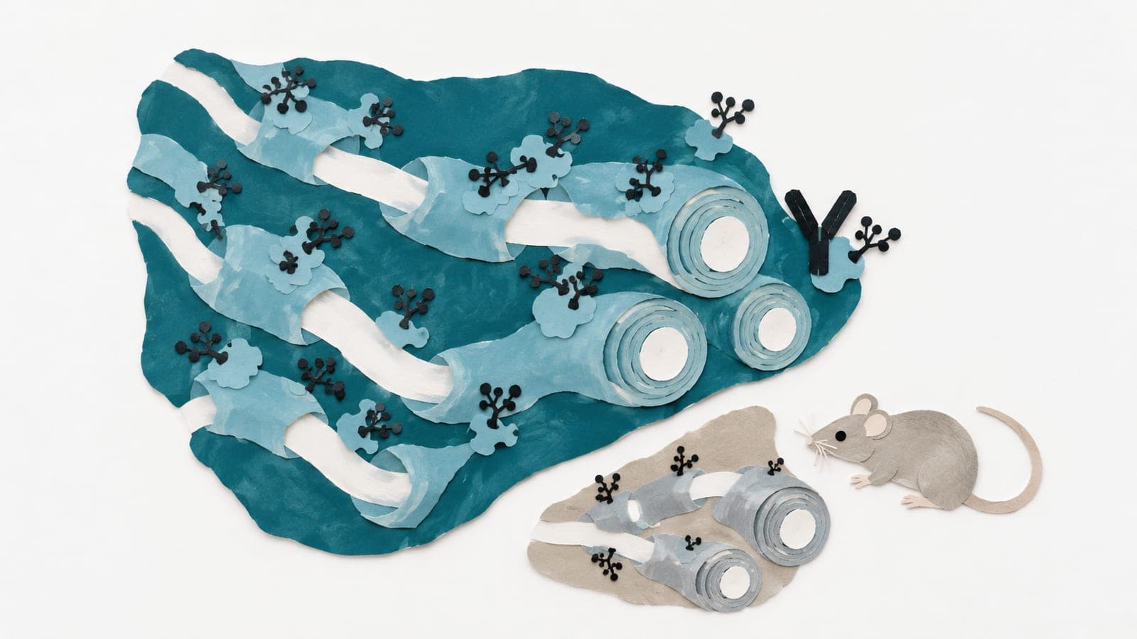
In mouse brains, short sugar chains called paucimannose increased with age, driven partly by an increase in an enzyme that processes sugars. In white matter, tissue rich in nerve fibers, aging increased attachment of these sugars to myelin oligodendrocyte glycoprotein, a target of immune attacks. Separately, researchers identified antibodies against this protein in older people and patients with an autoimmune disease involving such antibodies. Binding experiments showed that a sugar chain at one specific site on the protein altered antibody binding.
Why it matters
The work addresses how age-related changes in sugars attached to proteins may affect interactions with antibodies involved in brain autoimmunity.
Caveats
The aging atlas came from mice and does not establish how these patterns change across the human lifespan. The work is a preprint and has not been peer-reviewed.
Where the evidence stands
MOG
- 1 lab in 1 country
Immune response
- 293 independent labs in 46 countries
- 28 findings against
- Funders: National Institutes of Health and others
- 80 of 529 findings with company ties
- Resources: datasets, atlases and tools
- MOG
- Immune response
- MOG antibody-associated disease
- Experimental autoimmune encephalomyelitis
- Mice
The paper
Stanford University
bioRxiv, Oct 31, 2026, Open access, CC BY-NC-ND, Preprint, not peer-reviewed