Nuclear anchors weaken in senescent cultured human retinal cells
In a laboratory experiment, chemically induced senescent human retinal cells showed increased mobility and altered binding of proteins that anchor DNA to the nuclear scaffold.
bioRxiv
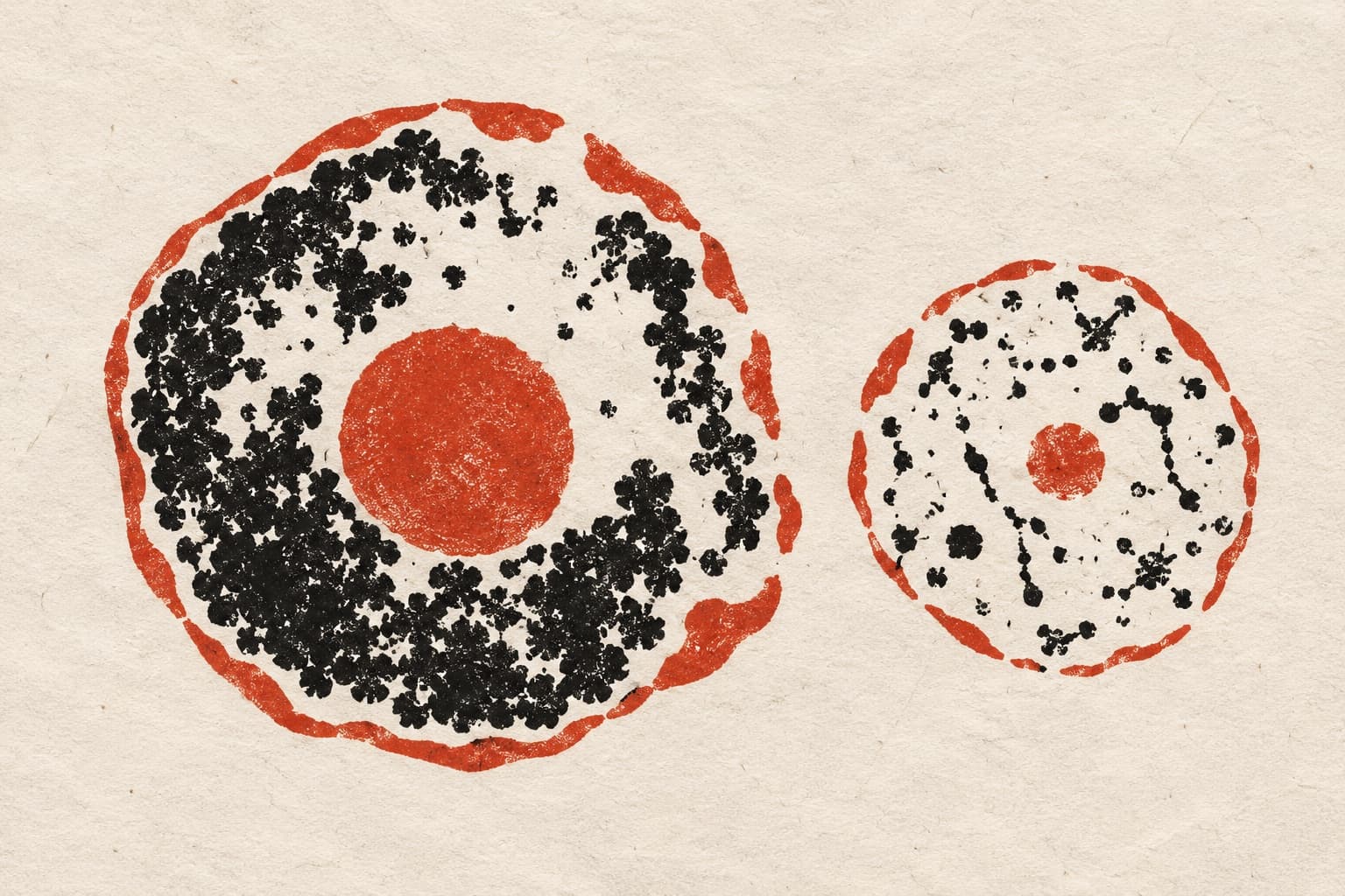
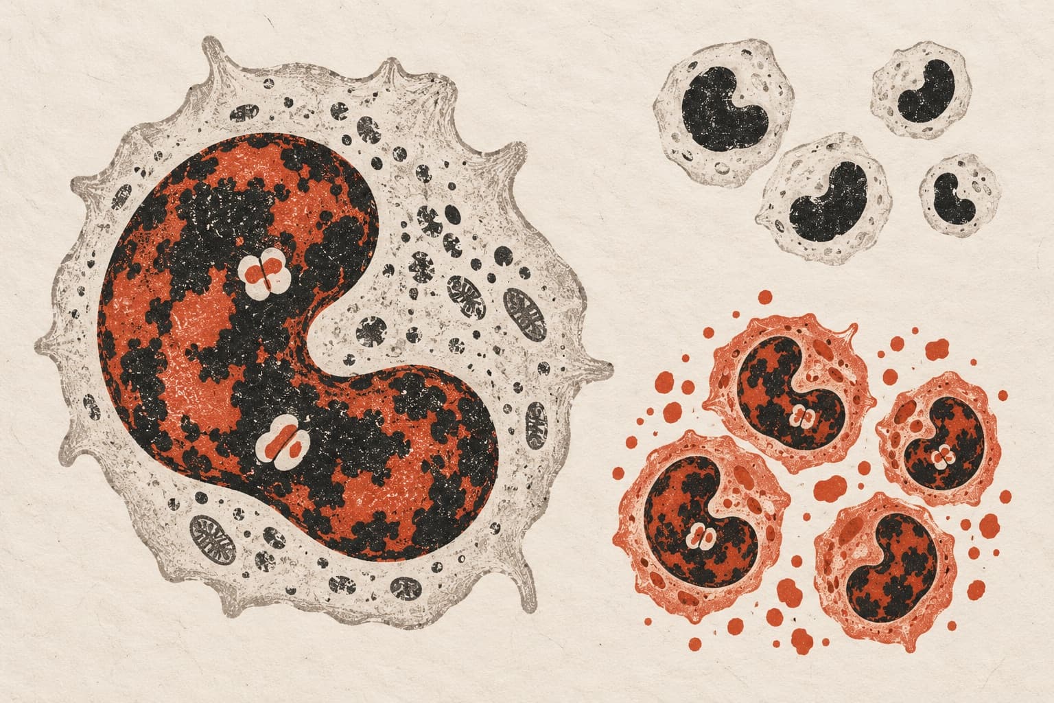
In an experiment in cells grown in the lab, researchers studied live human retinal pigment epithelium cells triggered into senescence, a state in which cells permanently stop dividing. The team examined the nuclear lamina, a structural scaffold that lines the cell nucleus. Senescent cells showed altered levels of two anchor proteins, lamin B receptor and emerin. Live-cell diffusion measurements showed that both proteins moved more freely, pointing to weakened anchoring to the nuclear scaffold. Single-molecule tracking also uncovered altered binding dynamics for each protein over space and time. In addition, dense domains of tightly packed DNA linked to regions of slowed lamin B receptor mobility became dispersed during senescence.
Why it matters
Restructuring of the nuclear scaffold is a defining feature of cell senescence, but its fine-scale physical behaviour remains poorly understood. The findings point to how loosened physical anchoring and rearranged DNA architecture could potentially drive changes in gene activity during senescence.
Caveats
The findings come from an experiment in cultured human cells using a chemical trigger to induce senescence, which may not fully reflect how cells behave in living tissue. The study was published as a preprint and has not yet been peer-reviewed.
The paper
National University of Singapore
bioRxiv · 9 Oct 2026 · CC BY-NC-ND · Preprint, not peer-reviewed