Chest scans helped detect hip osteoporosis in older adults
Chest-scan bone density at or below 85.4 milligrams per cubic centimeter helped identify hip osteoporosis when compared with standard hip scans.
Multicenter
Limit: The abstract reports no validation in an independent patient group
Edited by Boris Djordjevic · AgingResearch.News
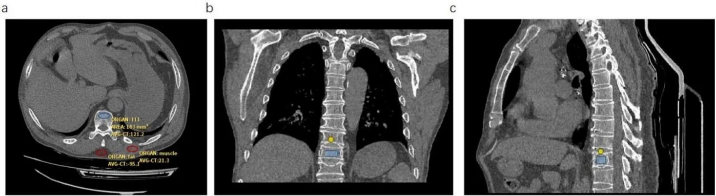
Researchers analyzed scans from 568 patients aged 60 and older in a retrospective observational study across several centers. All had chest computed tomography, which produces detailed X-ray images, and hip bone-density scans. The researchers measured bone mineral density in the spine’s chest region using images taken without contrast dye. They compared these measurements with the hip scans. Hip osteoporosis, a condition that weakens bones, affected 36.3% of patients.
At a threshold of 85.4 milligrams per cubic centimeter, sensitivity—the ability to identify people with hip osteoporosis—was 0.851. Specificity—the ability to identify those without it—was 0.864. The researchers checked the diagnostic model through internal validation, including 500 rounds of resampling the study data.
Why it matters
The work addresses whether images already collected during routine care can support screening for age-related bone loss. The authors propose using them to identify osteoporosis risk and help guide intervention.
Caveats
This retrospective study used internal validation; the abstract does not report testing in an independent patient group. The findings do not establish screening performance in adults under 60.
The paper
Fujian Medical University
European Journal of Radiology, Oct 11, 2026
