Chest scan model identified scan-defined osteoporosis in adults
In a separate test group of 2,490 adults, the model achieved a discrimination score of 0.993, where 1 represents perfect separation.
18,298 adults, independent replication, multicenter
Limit: Shared AI-QCT measurement errors in chest and lumbar bone density could inflate model accuracy
Edited by Boris Djordjevic · AgingResearch.News
An observational, cross-sectional study analyzed low-dose computed tomography (CT) scans from 18,298 adults at 134 Chinese health check-up centers. Researchers used bone mineral density, a measure of bone’s mineral content, from chest vertebrae to build models for identifying osteoporosis in the lower spine. An artificial intelligence system defined osteoporosis as average bone mineral density below 80 milligrams per cubic centimeter in two lower-spine vertebrae.
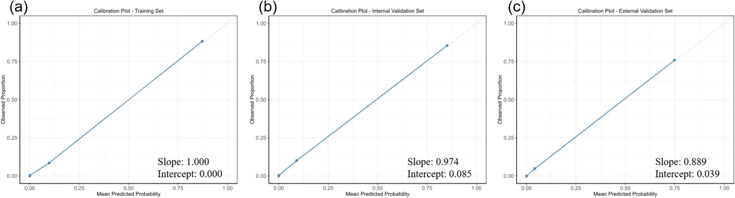
The team trained five models in 11,065 adults, then tested them internally in 4,743 and externally in 2,490. They selected logistic regression, a statistical model. In the external group, it achieved a discrimination score of 0.993; a score of 1 means perfect separation between people with and without the scan-defined condition. The authors suggested that adding this analysis to lung cancer screening may enable large-scale osteoporosis screening.
Why it matters
Osteoporosis is an age-related bone disease. The work addresses whether scans collected for lung cancer screening could also offer an opportunity to screen for osteoporosis.
Caveats
This cross-sectional study tested an artificial intelligence-defined scan outcome rather than osteoporosis validated against an independent diagnostic standard. The authors called for prospective validation against independent standards.


