Low-dose scans supported reliable brain protein measurements
Methods using computed tomography or magnetic resonance imaging agreed on positive brain protein scan results in 92.0–96.6% of cases.
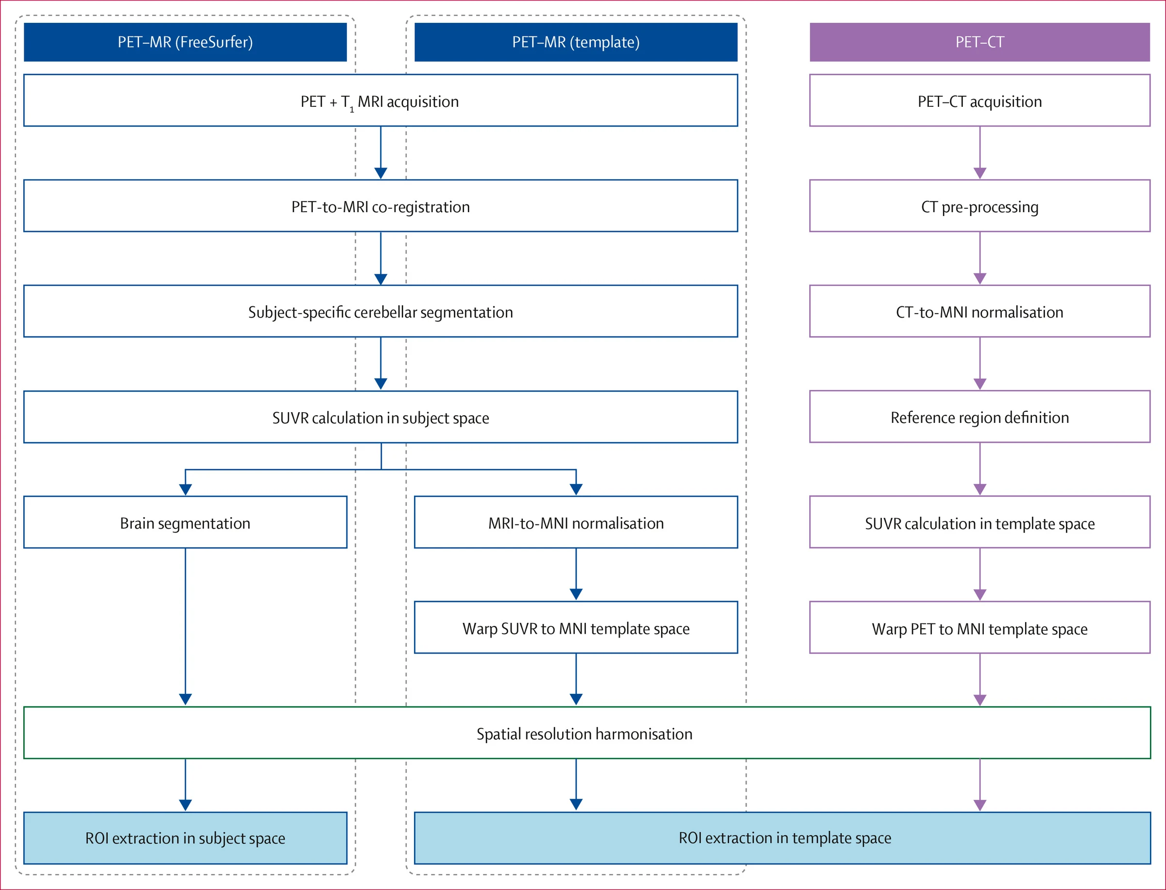
A six-site cross-sectional comparison studied 387 people aged 19–25, 50–65 or 66–89. Participants included 205 women and 182 men, ranging from people without thinking or memory impairment to those with mild impairment or dementia. Researchers compared ways to measure tau, a protein linked to Alzheimer's disease, using brain scans.
Participants underwent positron emission tomography, which uses radioactive tracers, alongside low-dose computed tomography and magnetic resonance imaging (MRI). Researchers tested whether the low-dose scans could replace MRI as the anatomical reference for calculating tau measurements. The resulting measurements closely matched those from MRI-based processing for both tau tracers tested. The methods also preserved associations between tau measurements and memory, and between tau measurements and amyloid, another protein associated with Alzheimer's disease.
Why it matters
For Alzheimer's disease research, using routinely collected low-dose scans could make datasets lacking MRI usable and broaden participation in clinical trials.
Caveats
This cross-sectional comparison assessed agreement between processing methods, not whether the approach improved diagnosis or patient outcomes. It did not assess whether low-dose computed tomography could replace MRI for other clinical purposes.
The paper
University of Pittsburgh
The Lancet Medical Imaging & Theranostics, 3 Aug 2026, Open access