Neck spinal cord size peaks in adulthood in lifespan charts
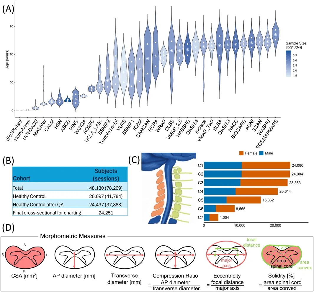
Modeled spinal cord area peaked around the mid-30s in a cross-sectional study of 24,251 healthy or cognitively normal people aged 0–100.
Limit: Spinal segments shift relative to vertebrae with age, potentially distorting apparent cord-size changes
Edited by Boris Djordjevic · AgingResearch.News
In a cross-sectional observational study, researchers analyzed brain scans from 24,251 healthy or cognitively normal people aged 0–100 across 33 datasets. They measured the size and shape of the spinal cord in the neck and built reference charts showing how these measures varied with age and sex.
The charts indicated rapid growth during childhood and adolescence, followed by peaks in early-to-mid adulthood and gradual decreases. Cross-sectional area—the area of a slice through the cord—peaked around the mid-30s. Size measurements also varied along the neck and were larger in males than females. The charts allow an individual's measurements to be compared with reference values for their age and sex.
Why it matters
Spinal cord measurements serve as indicators of neurological health. Age- and sex-specific references may help distinguish typical variation from changes linked to neurodegeneration.
Caveats
This cross-sectional study compared different people rather than following individuals over time, so its curves do not measure individual aging. The reference population consisted of healthy or cognitively normal controls.
From the paper
- Participants
This cohort included 67 participants: 34 controls (age 22.7–64.1; mean age 38.6; 26 female) and 33 participants with MS (age 25.0–64.4; mean age 42.4; 29 female).
- Limitation
First, our models are derived from cross-sectional data, which approximates lifespan trajectories but cannot capture individual longitudinal changes or potential cohort effects (Di Biase et al. 2023).
Quoted word for word from the full text on onlinelibrary.wiley.com.
- Modeling and theory
- Brain white matter volume
- Brainstem volume
- Cervical spinal cord anteroposterior dimension
- Cervical spinal cord AP/RL ratio
- Humans
The paper
Charting Cervical Spinal Cord Morphometry Across the Lifespan
Vanderbilt University
Human Brain Mapping, Oct 15, 2026, Open access