Brain scans trace three phases of nerve insulation with age
Across 214 brain regions in people aged 0–100, a scan measure rose rapidly in infancy, accelerated again in adolescence and adulthood, then declined late in life.
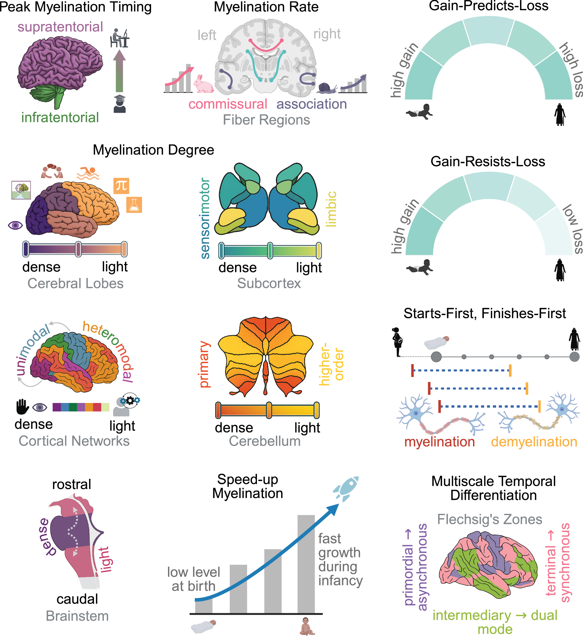
Limit: The MRI myelin marker also reflects iron and water content, not just nerve insulation
In people aged 0 to 100 years, researchers conducted an observational analysis of brain scans covering 214 regions. They used a ratio of two magnetic resonance imaging signals to estimate myelin, the insulating covering around nerve fibers.
The scan measure followed three phases across all regions: rapid growth in infancy, renewed acceleration in adolescence and adulthood, and decline in late life. The timing and patterns differed among brain regions. Researchers also examined regional brain volume and the thickness of the cortex, the brain’s outer layer, to assess how these structural features related to the myelin measure across life.
Why it matters
A reference for typical age-related myelin patterns may help researchers interpret changes linked to neurological disorders involving myelin.
Caveats
The observational analysis used a scan-based proxy for myelin rather than measuring it directly. The abstract did not specify the number of participants or whether the same people were followed over time.
From the paper
- Events by group
The largest increases in network-specific myelination occur between birth and infancy (Fig. 4D): (i) visual: 82.7%, (ii) dorsal attention: 78.8%, (iii) default mode: 73.5%, (iv) control: 72.5%, (v) somatomotor: 67.0%, (vi) salience attention: 66.2%, and (vii) limbic: 63.4%.
Quoted word for word from the full text on nature.com.
The paper
The shifting landscape of brain-wide myelination across the human lifespan
University of North Carolina at Chapel Hill
Nature Communications, Aug 29, 2026, Open access


