3D-bioprinted dual-factor nerve conduits promote neurovascular co-regeneration via activation of the PI3K/AKT and MAPK/ERK pathways
Abstract
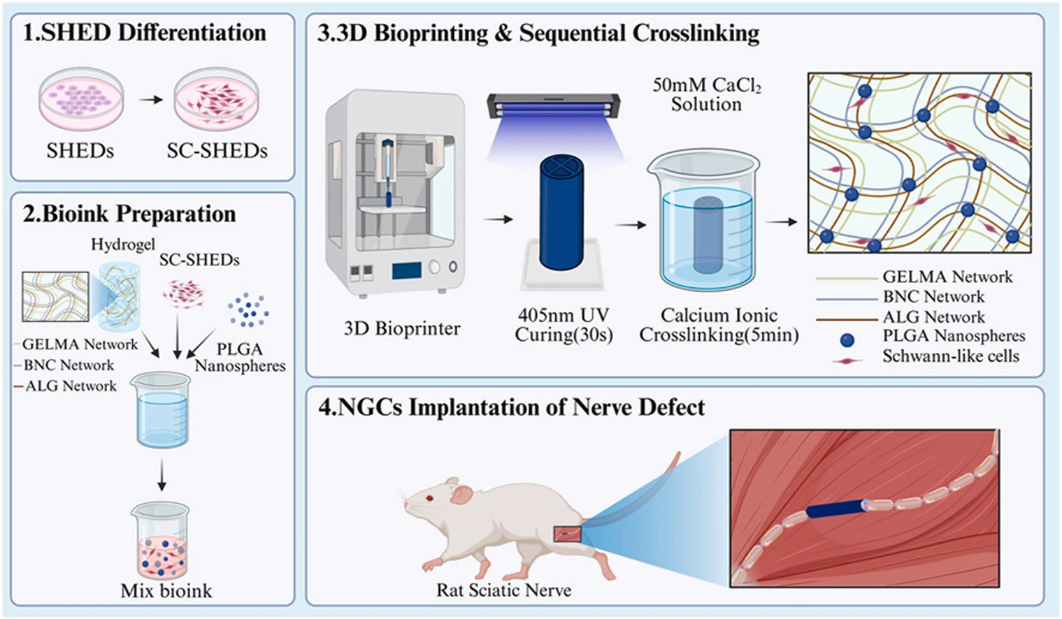
Peripheral nerve repair across challenging defects remains a major clinical hurdle, partly because conventional nerve guidance conduits fail to reconstruct the neurovascular microenvironment essential for coordinated regeneration. To overcome this, we engineered a 3D-bioprinted multichannel conduit composed of alginate (Alg), gelatin methacryloyl (GelMA), and bacterial nanocellulose (BNC), incorporating separately prepared nerve growth factor (NGF)- and vascular endothelial growth factor (VEGF)-loaded poly(lactic-co-glycolic acid) (PLGA) nanospheres and Schwann-like cells derived from stem cells from human exfoliated deciduous teeth (SC-SHEDs). The conduit was evaluated in a 10-mm rat sciatic nerve defect. Combined NGF/VEGF stimulation was associated with sustained AKT and ERK phosphorylation over the measured in vitro time course, together with changes in mTOR and c-Jun phosphorylation. The selected formulation retained the intended printed geometry, supported short-term cell viability, and enabled sustained release of both factors. Across the 1- and 3-month assessments, the NGF + VEGF + SC-SHED group showed increased microvessel density, stronger S100β- and NF200-associated signals, improved myelin morphometry, better electrophysiological and sensorimotor outcomes, and greater gastrocnemius muscle preservation than the untreated control. The combined conduit generally produced the most favorable outcomes among the engineered groups and approached the autograft group for several endpoints. These findings support further evaluation of this integrated strategy for peripheral nerve reconstruction.
- 3d-bioprinted dual-factor nerve conduit
- Nerve growth factor
- Angiogenesis
- MAPK/ERK signaling
- Peripheral nerve defect
- Rats
The paper
Sun Yat-sen University
Biomaterials, Oct 15, 2026

