Brain anatomy linked to proposed MRI marker in healthy adults
An observational study of 850 healthy young adults and 150 healthy aging participants linked MRI measurements to brain anatomy and the choice of brain regions analyzed.
850 participants
Limit: Deliberately retaining extreme ventricular volumes could inflate the strength of the anatomy–marker association
An observational study analyzed diffusion MRI scans from 850 healthy young adults and 150 healthy aging participants in the Human Connectome Project. The researcher examined an index proposed to reflect the function of the brain’s waste-clearing fluid system and spaces around blood vessels.
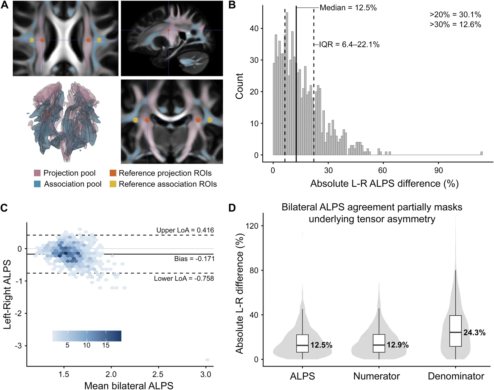
Standard measurements showed a median left-right difference of 12.5%. Refining the brain regions analyzed reduced measurement variability in independent validation groups, but differences between the brain’s two sides remained. Among healthy aging participants, the volume of the brain’s fluid-filled cavities explained substantially more variation in the index than age did. Lower index values were associated with expansion of these cavities and surrounding tissue. The findings suggest that the index combines anatomical influences rather than measuring brain fluid transport alone.
Why it matters
For research on healthy brain aging, a key question is whether this proposed marker can separate fluid-system function from anatomical differences.
Caveats
These were observational MRI measurements, not direct measurements of brain fluid transport. The participants were healthy, limiting conclusions about people with disease.
From the paper
- Limitation
First, no universally accepted DTI-ALPS ROI definition currently exists (Taoka et al., 2024).
Quoted word for word from the full text on direct.mit.edu.