Magnetic Particle Imaging of Macrophage Phagocytosis Evaluates Plaque Vulnerability and Therapeutic Efficacy in Atherosclerosis
Abstract
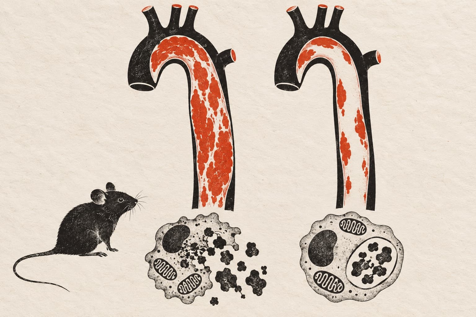
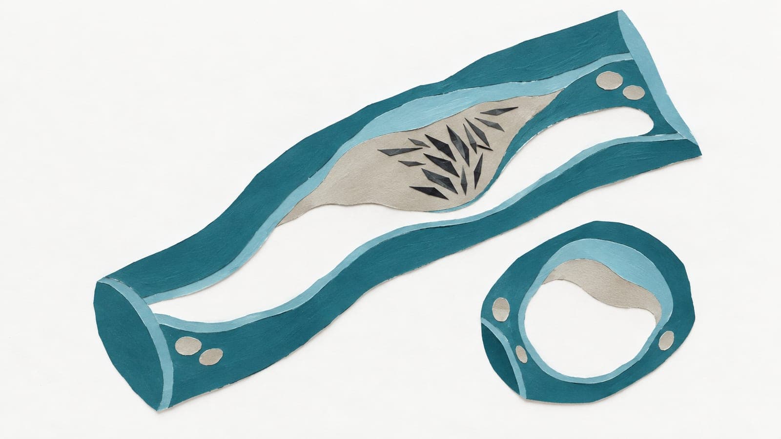
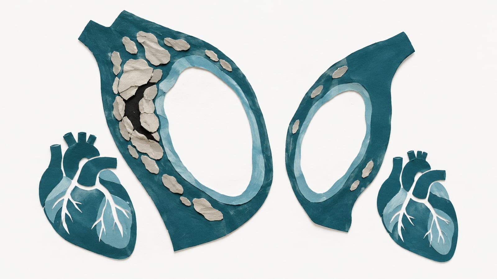
BACKGROUND: Developing new atherosclerosis therapeutics remains challenging. A noninvasive imaging method to assess plaque vulnerability is needed to provide early efficacy readouts and help select promising candidates for clinical trials. We aimed to identify macrophage phagocytosis markers associated with plaque vulnerability and evaluate whether magnetic particle imaging (MPI) of phagocytic activity can detect therapeutic responses and changes in plaque instability. METHODS: Integrated transcriptomic and immunofluorescence staining analyses of human carotid plaques were conducted to identify key phagocytosis-related markers. A novel polyethylene glycol-coated superparamagnetic iron oxide nanoparticle (PESPION) was synthesized and characterized alongside comparator agents (Vivotrax and ferumoxytol). Vulnerable plaques were induced in ApoE-/- mice by cast placement over the carotid artery. After 10-week treatment with a proprotein convertase subtilisin/kexin type 9 inhibitor (inclisiran) or a sodium-glucose cotransporter-2 inhibitor (empagliflozin), macrophage phagocytosis and plaque vulnerability were assessed by PESPION-enhanced MPI and histopathology, respectively. Comparisons between PESPION-enhanced MPI and magnetic resonance imaging were performed in proprotein convertase subtilisin/kexin type 9 inhibitor-treated and saline-control mice at 4 and 10 weeks. RESULTS: MSR1 (macrophage scavenger receptor 1) was identified as a phagocytosis marker associated with plaque vulnerability. Compared with Vivotrax and ferumoxytol, PESPION exhibited a larger core size, superior serum stability, enhanced MPI performance, and greater macrophage uptake, which was blocked by MSR1 inhibition. In vivo, PESPION-enhanced MPI sensitively detected vulnerable plaques, with signals reduced on MSR1 inhibition. Inclisiran and empagliflozin reduced MPI-quantified phagocytic activity compared with controls. MPI signals correlated positively with plaque area and macrophage infiltration, negatively with smooth muscle cell content, and predicted the vulnerability index. Direct comparison with magnetic resonance imaging revealed that MPI detected therapeutic responses earlier (4 weeks) than T2*-weighted magnetic resonance imaging (10 weeks). CONCLUSIONS: MSR1 is a phagocytosis-related molecule associated with plaque vulnerability. PESPION-enhanced MPI provides a sensitive, noninvasive tool for quantifying MSR1-mediated macrophage phagocytosis, enabling robust evaluation of plaque vulnerability and pharmacological response.
The paper
the Sixth Medical Center of Chinese PLA General Hospital
Arteriosclerosis, Thrombosis, and Vascular Biology, 15 Oct 2026