Varied follicle growth buffers egg cell lifespans in mice
Using lineage tracing across the lifespan of female mice, researchers mapped how secondary follicles regulate the survival of activated egg cells to support long-term fertility.
Nature Aging
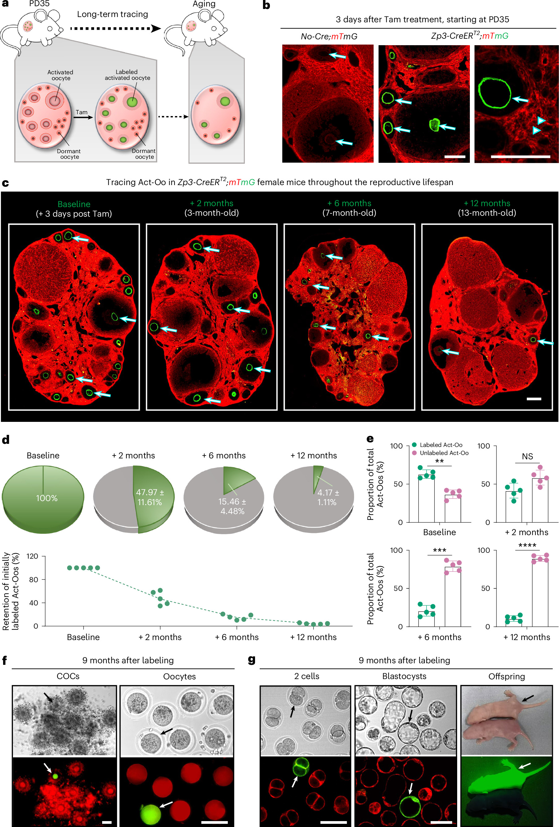
In female mice, researchers used a genetic tracking method to label activated egg cells and follow their development across the lifespan. The animal experiment showed that activated egg cells do not all mature at the same rate. Some eggs activated in early adulthood progressed rapidly, whereas others survived into late or even final reproductive stages, supporting long-term fertility. This lifespan was governed by the growth of secondary follicles, the structures of supporting cells that nurture developing eggs. The researchers found that secondary follicle progression acted as a buffer for reproductive capacity. Activated eggs displayed varied gene activity and metabolic states. Different levels of FURIN, a protein that processes other molecules, changed the release of chemical signals from the eggs, coordinating their development with surrounding follicles to regulate egg survival.
Why it matters
Oocyte activation and follicle growth are fundamental for mammalian fertility. Clarifying how tissues buffer the lifespan of activated egg cells bears on how female reproductive capacity is maintained across adult life.
Caveats
The findings come entirely from an experimental mouse model, and the timing and regulation of follicle development may differ in humans.
The paper
Show 13 more authors
Lingyu Li, Ge Wang, Lu Mu, Nan Sun, Kaiying Geng, Xindi Hu, Shudong Niu, Longzhong Jia, Yibo Wang, Xueqiang Xu, Zhen Li, Fengchao Wang, Yan Zhang,College of Biological Sciences
Nature Aging · 8 Oct 2026