Loss of myofibril number drives muscle fiber atrophy during aging and disuse
Structural analyses of humans and mice reveal that fibers shrink primarily by shedding myofibrils rather than reducing myofibril size.
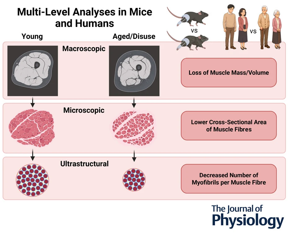
In young and older humans and mice, researchers examined the macroscopic to ultrastructural drivers of skeletal muscle loss. The team used magnetic resonance imaging to assess human quadriceps volume, alongside muscle biopsy imaging to measure fiber and myofibril characteristics. They also evaluated aged mice and a mouse model of disuse atrophy caused by unilateral limb immobilization. In humans, aging reduced muscle volume and cross-sectional area, causing radial atrophy in SERCA1-positive fibers while preserving SERCA2-positive fibers. This atrophy stemmed predominantly from having fewer myofibrils rather than smaller myofibrils. Mice showed similar aging patterns, though their SERCA1 myofibrils also shrank slightly. Disuse in mice likewise triggered muscle fiber atrophy driven almost entirely by a lower number of myofibrils.
Why it matters
Identifying myofibril loss as the conserved driver of fiber shrinkage provides a specific structural target for therapies aiming to counteract sarcopenia and disuse-related muscle wasting.
Caveats
The human findings rely on cross-sectional observations, and the disuse-induced atrophy experiments were limited to mice.
The paper
University of Wisconsin–Madison
The Journal of Physiology, Sep 25, 2026, Open access