Retinal imaging model predicts age and reveals sex differences in human aging
The retinal age gap correlates with mortality, disease risks, and sex-specific genetic pathways in more than 71,000 UK Biobank participants.
Nature Communications
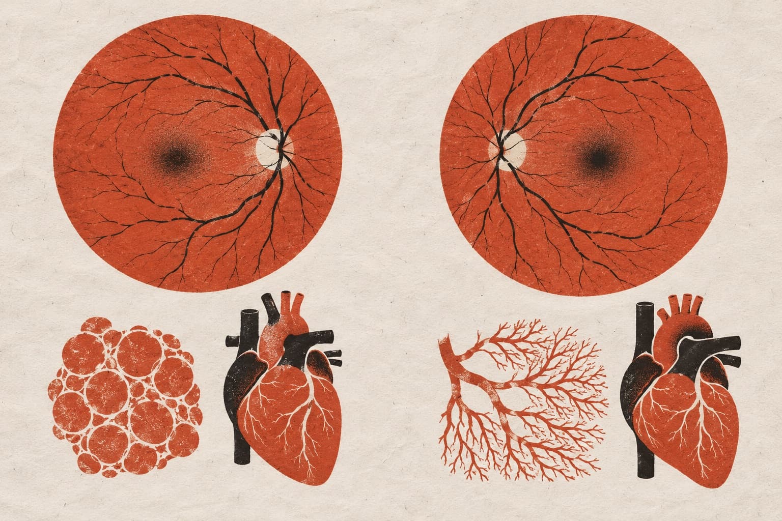
Researchers analyzed retinal fundus images from 71,343 human participants in the UK Biobank. They fine-tuned the deep learning foundation model RETFound to predict chronological age, reaching a mean absolute error of 2.85 years. The retinal age gap, defined as the difference between predicted and actual age, tracked with inflammation, cardiometabolic traits, cognitive performance, all-cause mortality, dementia, cancer, and incident cardiovascular disease. Genome-wide analyses linked this gap to genes involved in longevity, metabolism, neurodegeneration, and eye diseases. Sex-stratified analyses revealed distinct biological features. Retinal aging in males showed stronger ties to metabolic syndrome. In females, retinal aging involved vascular pathways and shifted across menopause, with postmenopausal women displaying higher age gaps and clinical profiles resembling those in males.
Why it matters
The findings indicate that non-invasive retinal imaging captures systemic aging processes and age-related disease risks. They also show that aging trajectories differ biologically between men and women.
Caveats
The study relied on observational data from a single cohort, the UK Biobank. It evaluated associations without directly demonstrating causal biological mechanisms.
The paper
Deep learning aging marker from retinal images unveils sex-specific clinical and genetic signatures
Show 21 more authors
Yating Pan, Bart J. Liefers, Jose D. Vargas-Quiros, Victor A. de Vries, Michael Johannes Beyeler, David M. Presby, Dennis Bontempi, Janna Hastings, Caroline C. W. Klaver, Ciara Bergin, Bogdan Draganski, Adham Elwakil, Györgyi Hamvas, Ilaria Iuliani, Ihor Kuras, Ilenia Meloni, Sofía Ortín Vela, Ian Quintas, Marc Schindewolf, Reinier O. Schlingemann, Mattia Tomasoni,SIB Swiss Institute of Bioinformatics
Nature Communications · 26 Aug 2026 · CC BY