Brain injury linked to white matter differences in women
A cross-sectional analysis of 363 women with recorded injuries and 10,128 controls linked brain scan differences to injury history and menopause timing.
Our read
Populations- Studied:
- Cross-sectional study of women with and without traumatic brain injury
- What changed:
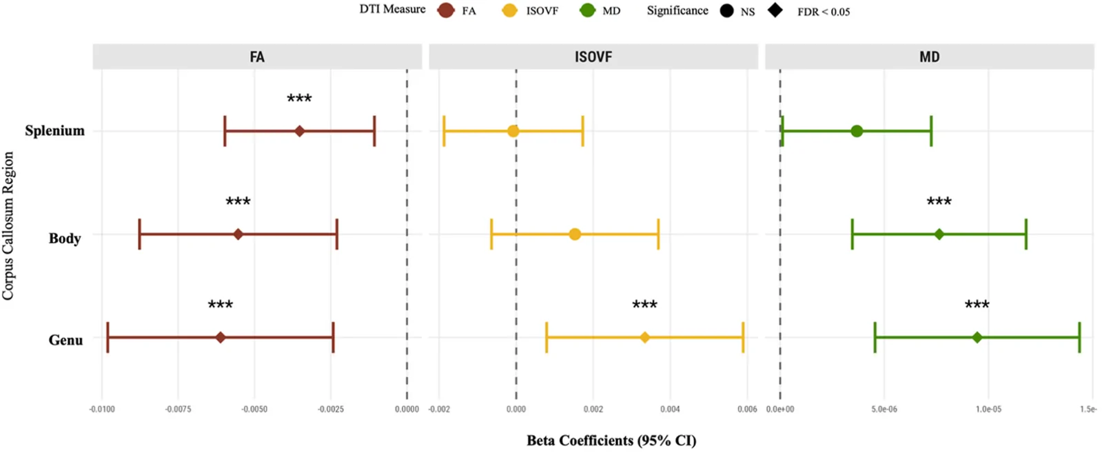
- Water diffusion in the corpus callosum body was higher in women with brain injury than in matched women without injury
- The catch:
- Primary analyses did not adjust for smoking, alcohol use, or physical activity
- In the record:
- The 417th paper on hormone replacement therapy with results in people; 909 on record in all.
- Why it’s aging research:
- It bears on staying healthy in midlife and later life.
Editor of record: Boris Djordjevic · AgingResearch.News
A cross-sectional study of female UK Biobank participants compared 363 women with recorded brain injuries within 10 years before imaging and 10,128 matched controls. Researchers examined the corpus callosum, a bundle of nerve fibers connecting the brain’s halves. Scan measures were available for 284 injury cases and 8,369 controls.
Women with injuries had scan patterns indicating less organized nerve fibers across several regions. The 30 women injured before menopause had lower scan measures of fiber organization in one region and greater water movement in another than women injured afterward and controls. Hormone replacement therapy use did not modify injury-related associations in the primary analyses. Among 3,108 hormone users, however, the association between duration of use and one scan measure differed between women with and without injuries.
Why it matters
Menopause timing offers a way to explore how hormonal history relates to brain structure after injury. The authors called for brain injury research that accounts for sex and hormonal exposure.
Caveats
Cross-sectional data cannot establish cause and effect, and 92.6% of injury cases were classified using broad head-injury codes, limiting conclusions about confirmed traumatic brain injury. The study excluded women who had undergone hysterectomy or removal of both ovaries.
From the paper
- Participants
The final analytical sample comprised 10,491 female participants, including 363 females with a history of TBI occurring 0-10 years prior to neuroimaging and 10,128 matched controls without TBI history.
- Result
Both reproductive factors independently predicted several corpus callosum measures: parity was associated with genu FA (standardized β=0.029, 95% CI [0.011, 0.048], p=0.002), body MD (standardized β=-0.053, 95% CI [-0.071, -0.034], p<0.001) …
- Limitation
First, UK Biobank participants are healthier and more educated than the general population, with this selection bias especially evident among those who completed neuroimaging.
Quoted word for word from the full text on PubMed Central.
- Hormone replacement therapy
- Sex hormone signaling (estrogen, androgen)
- Traumatic brain injury
- Fractional anisotropy
- Isotropic volume fraction
- Humans
The paper
Northeastern University
Women's Health, Sep 2, 2026, Open access
Comments
Be the first to comment.