Abdominal CT-derived metabolic phenotypes are associated with the MRI-derived brain age gap in women
Declared no competing interests
Abstract
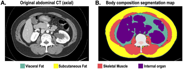
BACKGROUND: Metabolic dysfunction is increasingly recognized as a systemic contributor to adverse brain aging; however, conventional clinical markers may not fully capture tissue-level variations in liver attenuation, abdominal adiposity, and muscle quality. We examined whether abdominal CT-derived metabolic phenotypes were associated with MRI-derived brain age gap (BAG) in women. METHODS: This retrospective health-screening cohort included 1,280 women who underwent both abdominal CT and brain MRI between 2019 and 2024. Predicted brain age was estimated using brainageR, and the BAG was calculated as predicted brain age minus chronological age. CT-derived phenotypes were extracted from dual-energy CT virtual non-contrast images using DeepCatch software. The primary CT phenotypes were FatRatio, defined as the abdominal fat area relative to the fat plus skeletal muscle area, and liver-to-spleen attenuation ratio. RESULTS: In models adjusted for age, parity, body mass index, hemoglobin A1c, triglycerides, high-density lipoprotein (HDL) cholesterol, smoking, and alcohol use, a higher FatRatio was associated with a larger BAG (β = 0.108, p = 0.007), whereas a higher liver-to-spleen attenuation ratio was associated with a lower BAG (β = -0.123, p < 0.001). In joint models including both primary CT phenotypes and the same clinical covariates, FatRatio (β = 0.095, p = 0.037) and liver-to-spleen attenuation ratio (β = -0.115, p = 0.001) retained independent associations. These findings persisted after age-bias correction of BAG and exclusion of extreme BAG outliers. CT-derived markers added modest but significant explanatory value beyond conventional clinical covariates. CONCLUSION: These findings suggest that abdominal CT-derived liver attenuation and relative adiposity phenotypes may capture systemic tissue-level metabolic variations relevant to MRI-derived brain aging in women.
The paper
Seoul National University
Frontiers in Aging Neuroscience, 4 Aug 2026