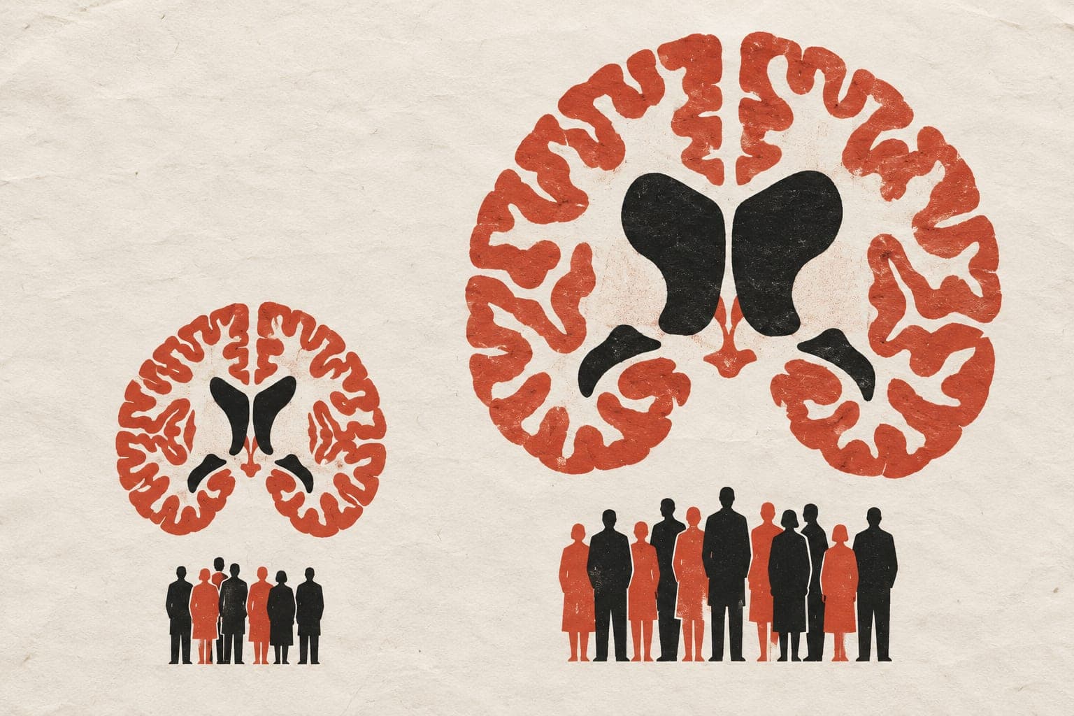
Brain regions reach their structural peak at different ages
In an observational study of 2,796 adults, a multimodal brain-age model predicted chronological age with an average error of 3.45 years.
Nature Communications
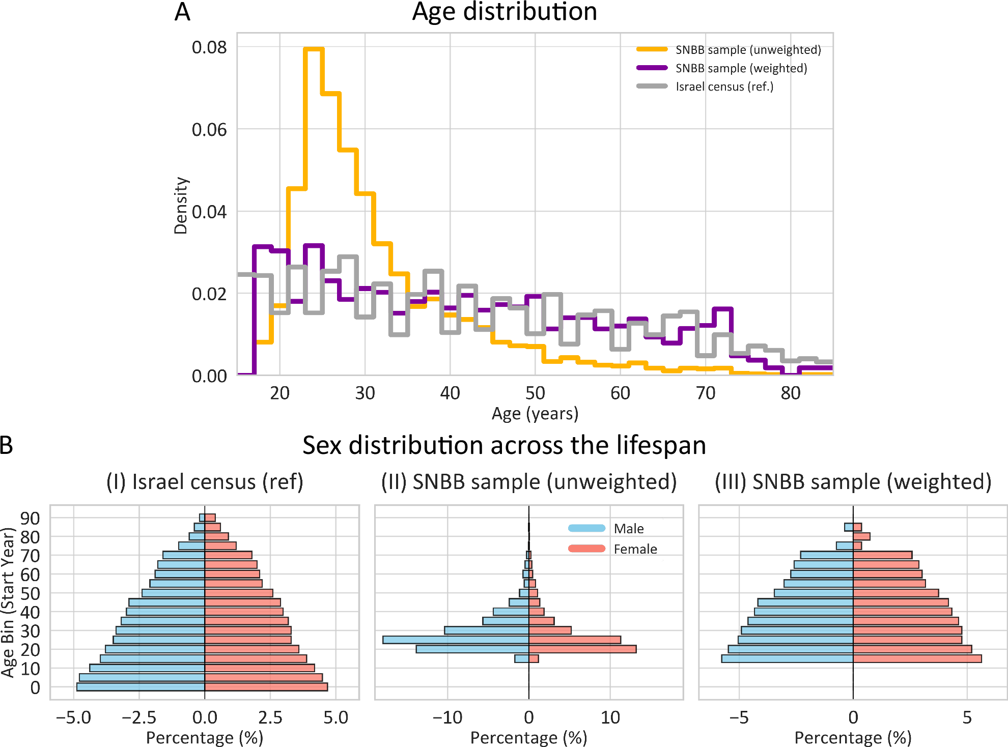
In an observational study of 2,796 adults, researchers analysed brain scans to map how different brain regions change across adult life. The team combined measures of grey-matter volume with diffusion scans, which track tissue microstructure. They identified an age of structural transition for each region, marking maximal structural refinement before age-related reduction. Sensory and motor cortex peaked early, whereas association networks peaked later. Distant brain regions grouped into networks sharing multimodal aging trends rather than anatomical proximity. Combining regional patterns predicted chronological age with a mean absolute error of 3.45 years. Brain-age gaps showed links to phenotypes, or observable traits, that were weak in younger adults and amplified later in life.
Why it matters
Brain aging is often treated as uniform across the whole organ. Mapping when distinct networks peak and decline provides a framework for exploring how varied structural changes relate to individual traits across the lifespan.
Caveats
Transition ages were derived from statistical models fitted to age associations rather than direct physical observations of aging. The study was observational in 2,796 adults, showing associations rather than causal mechanisms.
- Machine learning
- Age of Structural Transition
- Brain age gap
- Gray matter volume
- Regional timing of structural transition
- Humans
The paper
Divergent Multimodal Age-Association Profiles Across the Human Brain
Tel Aviv University
Nature Communications · 9 Sep 2026