Senescent cell secretions injure lungs in newborn mice
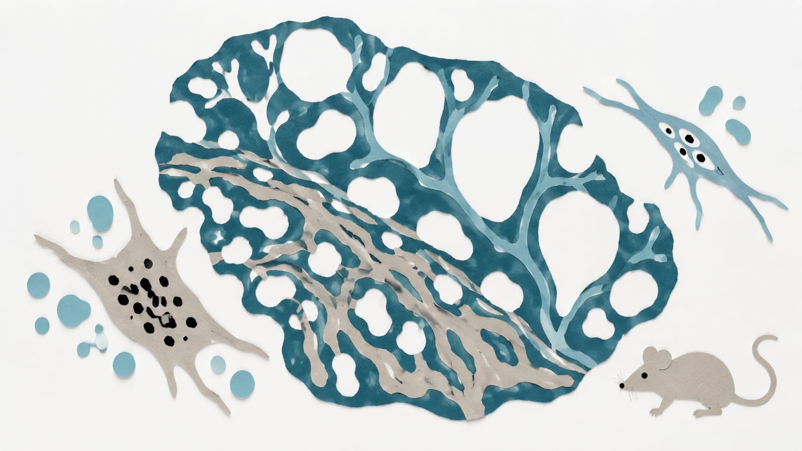
Secretions from oxygen-exposed mouse lung immune cells disrupted air sac and blood vessel development in newborn mice assessed 14 days after birth.
Our read
Mechanisms- Studied:
- Experiments in newborn mice
- What changed:
- Fluid from senescent cells caused simpler air sac and blood vessel structures in newborn mice
- The catch:
- The inhibitor may have remained in transferred fluid and acted after transfer
Editor of record: Boris Djordjevic · AgingResearch.News
Experiments in newborn mice and mouse lung immune cells grown in the lab examined how high oxygen exposure contributed to lung injury. In mouse lung data, high oxygen increased markers of senescence, a state in which cells stop dividing, in macrophages, immune cells in the air sacs. In cultured cells, 24 hours of high oxygen increased sugar breakdown for energy. Inhibiting this process reduced senescence markers. High oxygen also reduced cell movement and the ability to engulf beads.
Researchers gave newborn mice fluid from the cultured cells on days 7 and 10 after birth and examined their lungs on day 14. Fluid from senescent cells caused simpler air sac and blood vessel structures. Fluid from oxygen-exposed cells caused less injury when the cells had received a drug blocking p38, a protein that relays signals within cells.
Why it matters
Senescence is an aging-related process that may also contribute to injury in developing lungs. The authors propose targeting macrophage sugar metabolism and p38 signaling to prevent lung injury.
Caveats
The inhibitor may have remained in the transferred fluid, so researchers could not determine whether it acted only on the macrophages or also after transfer. These were mouse and cell-line experiments, not tests in premature infants.
- Senescent and/or hyperoxia-conditioned media
- Cellular senescence
- Glycolysis
- Lung injury
- Senescence markers
- Mice
The paper
Neonatal hyperoxia promotes macrophage senescence and lung injury via a p38MAPK-dependent secretome
Brown University
American Journal of Respiratory Cell and Molecular Biology, Jul 17, 2026, Open access
Comments
Be the first to comment.