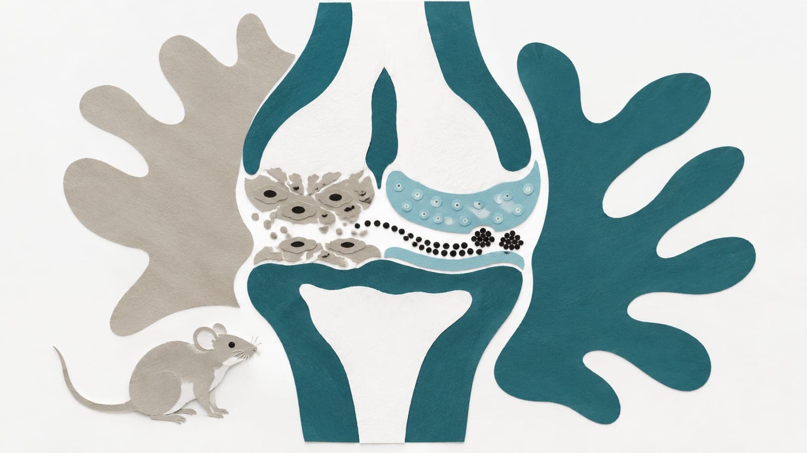
Structural and functional changes in articulating joints during aging of male and female C57BL/6JN mice
Abstract
OBJECTIVE: Define the natural history of articular joint changes in aging C57BL/6JN mice. DESIGN: Male and female C57BL/6JN mice of various ages were obtained from the National Institute on Aging (NIA). Knee joints were examined with atomic force microscopy (AFM), histology, and for Proteoglycan 4 (Prg4) mRNA expression and compared across different ages for each sex. RESULTS: In male mice, histological damage on tibiae (p = 0.04; 95% CI: -41.3, -0.88) and elastic moduli of femoral middle zone articular cartilage (p= 0.04; 95% CI -1.83, -0.03) increased between 12 and 16 months, while Prg4 expression in the superficial zone decreased (p=0.02; 95% CI: 0.02, 0.38). No aged-related changes in tissue histology or Prg4 expression were detected in the superficial zone of articular cartilage in female C57BL/6JN mice; however, increases in elastic moduli were apparent in the middle zone from 16 to 25 months (p=0.002, 95% CI -2.77, -0.72). Meniscal pathology was not observed in female mice at any age and was prevalent in 27-m/o males (p= 0.0005; 95%CI: -11.75, -2.61). CONCLUSIONS: Male C57BL/6JN mice from the NIA aging rodent colony develop signs of cartilage degeneration between 12 and 16 months of age in several assays. AFM on the middle zone of articular cartilage is a sensitive method to detect degeneration in male and female mice and is the only assay to detect changes in female articular cartilage at 16 months of age. These results inform the design of therapeutic strategies for joint diseases in this mouse strain.
The paper
Mayo Clinic
Osteoarthritis and Cartilage, Jun 12, 2026, Open access