Modified stem cells promote blood vessel formation in mice
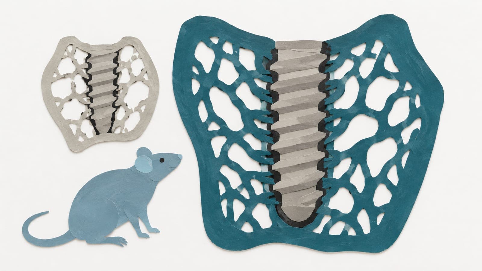
At 14 days, cells penetrated 591.0 micrometers into modified-cell scaffolds implanted in mice, versus 377.3 micrometers in control scaffolds.
Our read
Interventions- Studied:
- Experiments in 16 Nude CBA mice aged 9–10 weeks, 28 days, against controls
- What changed:
- Blood vessels covered more scaffold area and more vessels were larger in modified-cell scaffolds than in controls
- The catch:
- Fracture healing and aging were not measured
- Why it’s aging research:
- It studies how the body can be repaired or renewed.
Editor of record: Boris Djordjevic · AgingResearch.News
In 16 Nude CBA mice aged 9–10 weeks, researchers tested human bone marrow-derived mesenchymal stem cells, which can form bone and other tissues. The animal experiment used scaffolds made of collagen and a bone mineral, implanted under the skin. Each mouse received modified cells on one scaffold and control cells on another. The modification inhibited miR-195, a small RNA that regulates gene activity.
At 14 days, cells had moved deeper into scaffolds carrying modified cells. These could include both implanted human cells and cells from the mice. At 28 days, modified-cell scaffolds had more collagen and higher activity of genes associated with bone formation. Blood vessels covered more scaffold area, and more vessels fell into the larger-size category, than in controls.
Why it matters
Bone repair matters for skeletal health as people age. The authors suggest that engineering stem cells may improve cell-based approaches for musculoskeletal diseases and bone defects.
Caveats
This was an implant experiment in young mice, not a test of fracture healing or aging. Two mice with scaffold-associated infections were excluded at day 28, and incomplete tissue recovery excluded another mouse’s samples at day 14.
The paper
Universidade do Porto
Stem Cell Research & Therapy, Jun 11, 2026, Open access
Comments
Be the first to comment.