Type 2A fibers were richer in mitochondria in mouse muscles
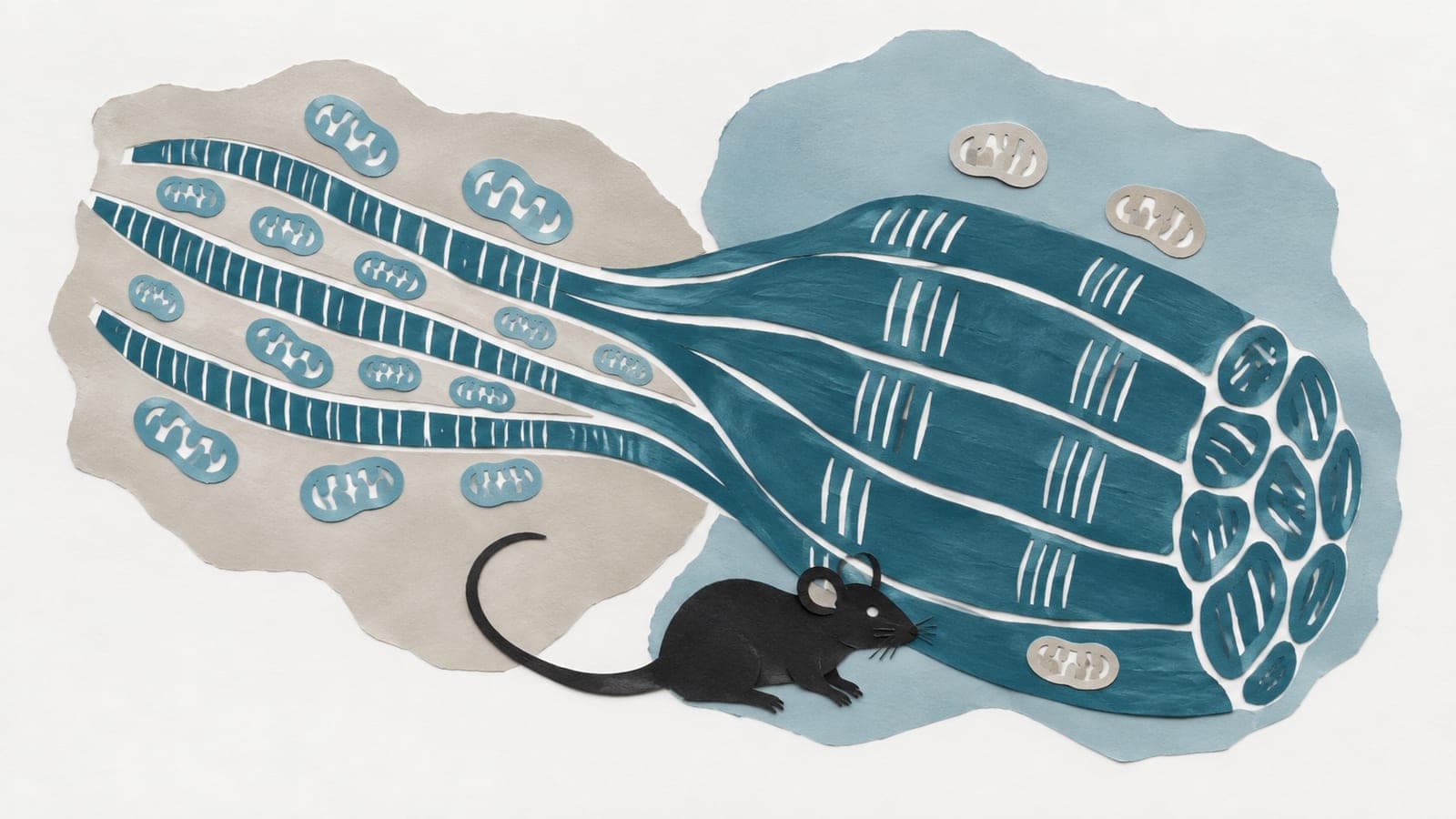
Experiments across mouse muscle regions found higher mitochondrial content in type 2A than type 1 fibers, a pattern maintained during aging and exercise.
Our read
Mechanisms- Studied:
- Experiments in mice, against type 1 fibers
- What changed:
- Type 2A fibers had higher mitochondrial content than type 1 fibers across different skeletal muscle regions in mice
- The catch:
- The abstract does not report mouse numbers, ages, or exercise conditions
- In the record:
- One of 9,759 papers on exercise on record: 3,810 with results in people, 603 in animals. Its result on mitochondrial content agrees with all 4 other findings on record.
- Why it’s aging research:
- It studies an intervention with evidence of extending healthy life.
Editor of record: Boris Djordjevic · AgingResearch.News
In experiments in mice, researchers examined mitochondrial content in individual skeletal muscle fibers across multiple muscle regions. Muscle fibers are muscle cells, and mitochondria are structures that supply energy. The team investigated how mitochondrial content differed among fiber types under physiological conditions including aging and exercise.
Fast-twitch type 2A fibers had higher mitochondrial content than slow-twitch type 1 fibers across the muscle regions examined. This relationship persisted under aging and exercise conditions. The researchers also identified type 2XD fibers, another fast-twitch type, as a group that responded dynamically to changes in physiological conditions.
Why it matters
Muscle cells adjust mitochondrial content to meet energy demands. The work bears on how those adjustments differ among muscle fiber types during aging.
Caveats
These were experiments in mice, not people. The abstract does not report the number or ages of the mice or details of the exercise conditions.
- Exercise
- Mitochondrial biogenesis (PGC-1α)
- Mitochondrion
- Mitochondrial content
- Mitochondrial enzyme activities
- Mice
Exercise is part of Bryan Johnson's Blueprint
The paper
Tokyo Medical University
Biochemical and Biophysical Research Communications, May 8, 2026
Comments
Be the first to comment.