Protective effect and mechanism of TSPAN9-mediated mitocytosis in interleukin-1β-induced rat chondrocyte senescence
Abstract
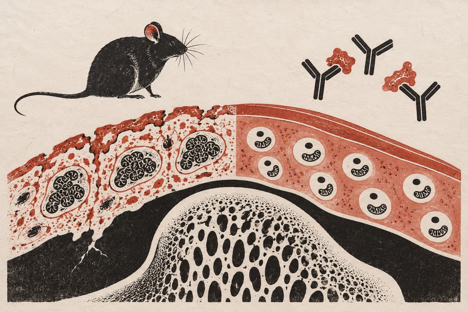
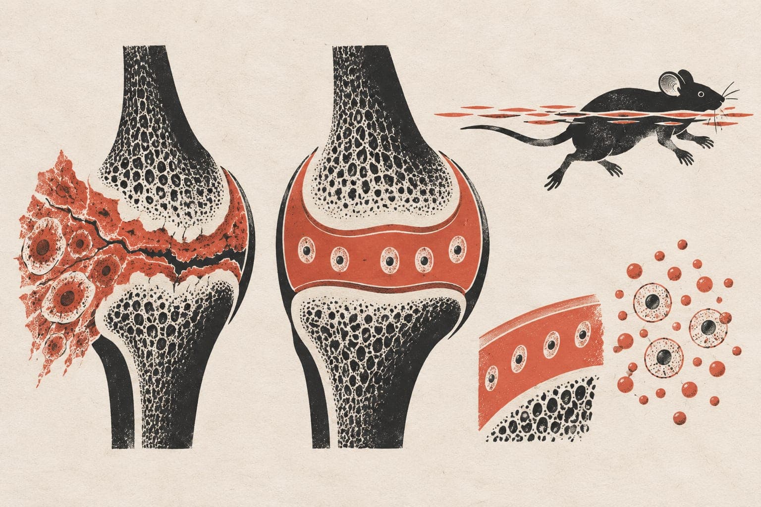
OBJECTIVE: To investigate the regulatory role and molecular mechanisms of TSPAN9-mediated mitocytosis in an interleukin-1β (IL-1β)-induced rat chondrocyte senescence model, and to identify novel therapeutic targets for osteoarthritis (OA). METHODS: Primary knee articular chondrocytes were isolated from the cartilage of knee joints harvested from 1-week-old male Sprague-Dawley rats and maintained in culture. Cell viability was assessed using the cell counting kit 8 (CCK-8) assay, and cell-cycle distribution was analyzed by flow cytometry to determine the optimal concentration and exposure time of IL-1β for model induction, thereby establishing an in vitro chondrocyte senescence model [early-OA (E-OA), middle-OA (M-OA), late-OA (L-OA)] and grouped. Cellular senescence was evaluated by senescence-associated β-galactosidase (SA-β-gal) staining. Real-time quantitative PCR (qRT-PCR) was used to quantify the mRNA expression levels of senescence markers [cyclin-dependent kinase inhibitor 2A (Cdkn2a) and Cdkn1a], extracellular matrix (ECM) catabolic-anabolic genes [collagen type Ⅱ alpha 1 chain (Col2a1), Aggrecan (Acan), matrix metalloproteinase 13 (MMP-13), and a disintegrin and metalloproteinase with thrombospondin motifs 5 (ADAMTS5)], and key mitocytosis-related genes [kinesin family member 5B (KIF5B), TSPAN9, and TSPAN4]. Mitochondrial ultrastructure and mitocytosis-related morphological features were examined by transmission electron microscopy, and changes in mitochondrial membrane potential were assessed using the JC-1 fluorescent probe. To activate TSPAN9 expression, chondrocytes were transduced with a TSPAN9-overexpressing lentivirus (LV-TSPAN9). The experiments included four groups: control group, M-OA group, lentivirus negative control group, and LV-TSPAN9 group. After confirming overexpression efficiency, differences in cellular senescence, mitochondrial homeostasis, and key ECM metabolic indices were compared among the groups. RESULTS: Using CCK-8 assays and cell-cycle analysis, the staged rat chondrocyte senescence model induced by IL-1β (5 ng/mL), and defined 12, 24, and 48 hours as the E-OA, M-OA, and L-OA senescence phenotypes were successfully established, respectively. Model validation demonstrated that, with prolonged induction, the proportion of SA-β-gal-positive cells increased markedly, mitochondrial membrane potential progressively declined ( P<0.05), and mitochondrial ultrastructural damage became increasingly severe. qRT-PCR showed progressive upregulation of the senescence markers Cdkn2a and Cdkn1a, as well as the ECM catabolic genes MMP-13 and ADAMTS5 ( P<0.05), together with sustained downregulation of the ECM anabolic genes Col2a1 and Acan ( P<0.05). Notably, the key mitocytosis-related genes KIF5B, TSPAN4, and TSPAN9 displayed a biphasic pattern, with early compensatory upregulation followed by decompensatory decline at the middle-to-late stages. In the M-OA model, TSPAN9 overexpression markedly reversed these pathological changes, restored mitochondrial membrane potential ( P<0.05), ameliorated the senescent phenotype and ECM metabolic imbalance, and significantly upregulated the expression of mitocytosis-related genes and proteins ( P<0.05), with transmission electron microscope revealing morphological structures suggestive of mitocytosis-related events. CONCLUSION: Impaired mitocytosis is an important pathological mechanism underlying IL-1β-induced chondrocyte senescence, and TSPAN9 overexpression may delay chondrocyte senescence by promoting mitocytosis in concert with KIF5B, thereby facilitating the clearance of damaged mitochondria and restoring intracellular homeostasis.
The paper
Sichuan University
Zhongguo Xiu Fu Chong Jian Wai Ke Za Zhi, Apr 15, 2026