Old Age Drives Fusion and Expansion of Vitellogenin Vesicles in the Intestine of Caenorhabditis elegans
Abstract
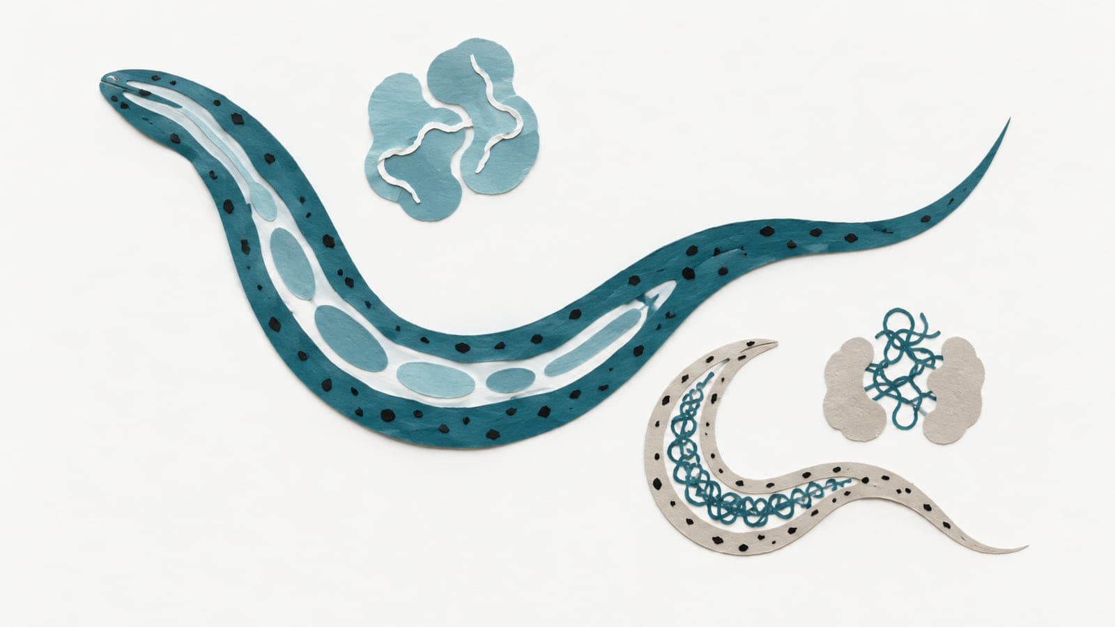
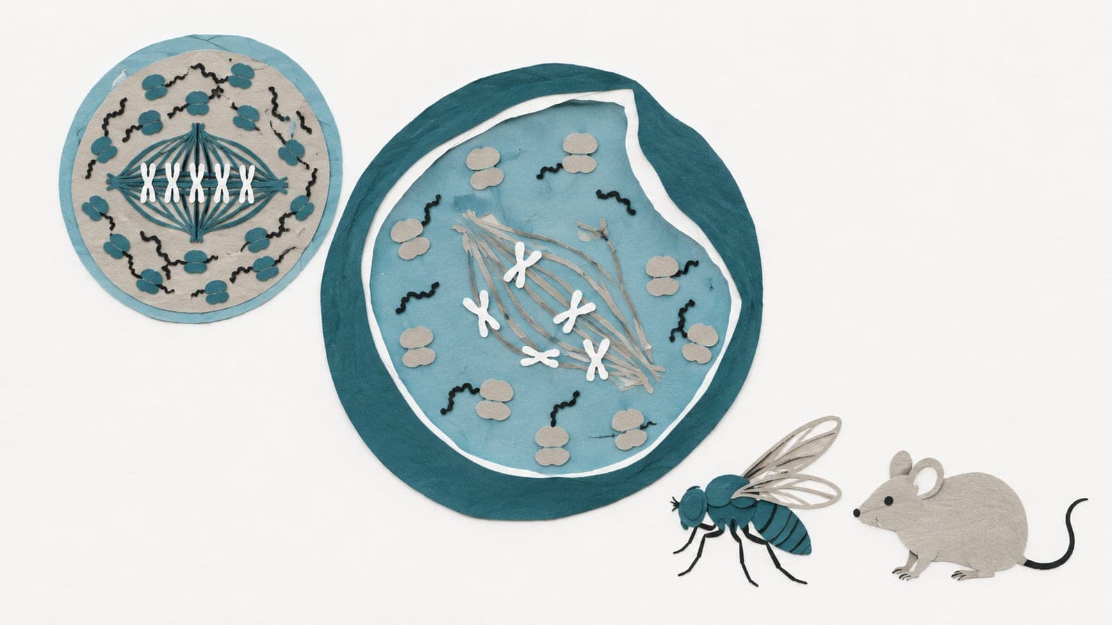
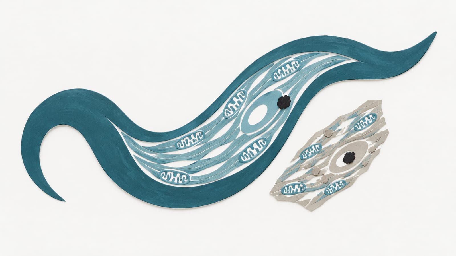
Some of the most conspicuous aging phenotypes of C. elegans are related to post-reproductive production of vitellogenins (Vtg), which form yolk protein (YP) complexes after processing and lipid loading. Vtg/YP levels show huge increases with age, and inhibition of this extends lifespan, but how subcellular and organism-wide distribution of these proteins changes with age has not been systematically explored. Here this has been done to understand how vitellogenesis promotes aging. The age-associated changes of intestinal vitellogenin vesicles (VVs), pseudocoelomic yolk patches (PYPs), and gonadal yolk organelles (YOs) have been characterized by immuno-electron microscopy. We find that from reproductive adult day 2 (AD 2) to post-reproductive AD 6 and AD 9, intestinal VVs expand from 0.2 μm to 3-4 μm in diameter or by > 3,000 times in volume, PYPs increase by > 3 times in YP concentration and volume, while YOs in oocytes shrink slightly from 0.5 μm to 0.4 μm in diameter or by 60% in volume. In AD 6 and AD 9 worms, mis-localized YOs found in the hypodermis, uterine cells, and the somatic gonadal sheath can reach a size of 10 μm across in the former two tissues. This remarkable size increase of VVs and that of mis-localized YOs in post-reproductive worms are accompanied by extensive fusion between these Vtg/YP-containing vesicular structures in somatic cells. In contrast, no fusion is seen between YOs in oocytes. We propose that in addition to the continued production of Vtg, excessive fusion between VVs and mislocalized YOs in the soma worsen the aging pathologies seen in C. elegans .
- Intestinal vitellogenin vesicle size
- Pseudocoelomic yolk patch volume
- Yolk organelle size
- Yolk protein concentration
- C. elegans
The paper
Peking University; National Institute of Biological Sciences, Beijing
bioRxiv, Jul 22, 2022, Open access, CC BY-NC-ND, Preprint, not peer-reviewed