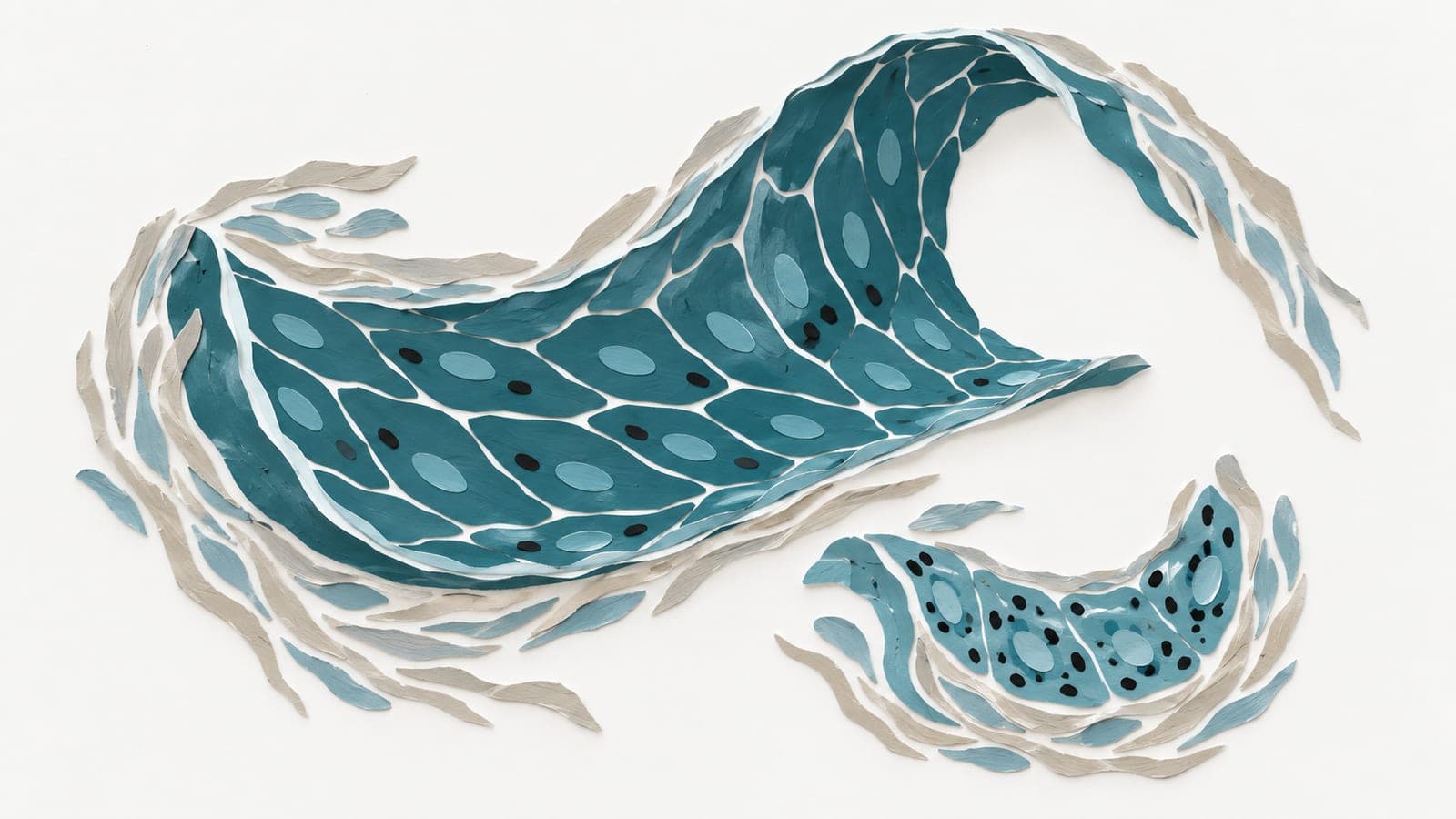
Heart electrical fibers remodel and fragment with age in sheep
Comparing heart tissue from sheep identified 407 genes enriched in electrical conduction fibers relative to surrounding muscle and sheath tissue, with bundles fragmenting in older hearts.
Limit: Fiber changes within individual sheep over time were not measured
Edited by Boris Djordjevic · AgingResearch.News
In an experiment using heart tissue from young (1 year) and mature (older than 6 years) sheep, researchers isolated Purkinje fibers, cells that conduct electrical impulses through the heart. Using laser microdissection, they compared these fibers with adjacent heart muscle and surrounding sheath tissue. The comparison identified 407 genes consistently more active in Purkinje fibers than in muscle and sheath tissue across both age groups. From these, the researchers prioritized 10 candidate genes. Laboratory tests in isolated sheep fiber strands confirmed that 9 of the 10 candidates showed 6- to 900-fold enrichment compared to heart muscle. Protein staining showed conserved enrichment patterns in human ventricular tissue samples.
In mature sheep hearts, genes for cell adhesion and mitochondria, the cell's energy producers, showed coordinated lower activity. Microscopic analysis revealed increased partitioning by collagen, a structural protein, and fragmentation of fiber bundles.
Why it matters
Dangerous ventricular arrhythmias, or irregular heartbeats, become more frequent with advancing age, and the Purkinje network can trigger these abnormal rhythms. Mapping how these conduction fibers change structurally and molecularly provides a biological foundation for studying why aging hearts become prone to rhythm disorders.
Caveats
The findings come from cross-sectional comparisons rather than tracking individual animals over time, and the structural aging changes were observed in sheep tissue rather than living people. In addition, the results were reported in a conference abstract, and human tests were limited to tissue samples.
- Transcriptomics
- Extracellular matrix remodeling
- Extracellular matrix
- Adhesion and mitochondrial transcript expression
- Collagen partitioning
- Sheep